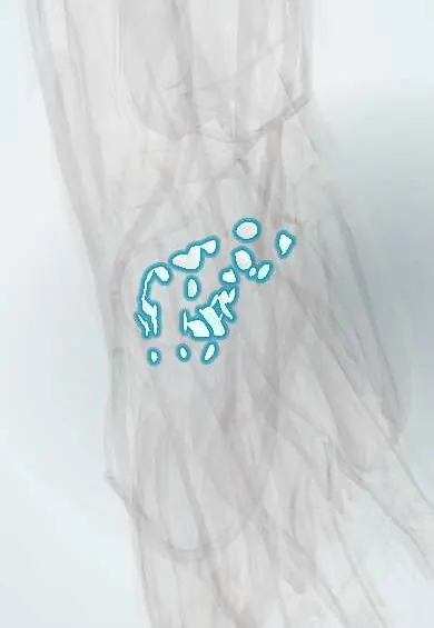
Joints and ligaments · Upper limb
CMC ligaments !
The carpometacarpal ligaments join the wrist bones to the palm bones, making the palm a stable arch; the thumb's are looser so it can move.
Official name: Carpometacarpal and metacarpal ligaments
Opens in the Durgical app, no sign-up needed.
What it is
Ligaments joining the wrist bones to the bases of the metacarpals and the metacarpals to each other.
What it does
Make the palm a stable arch; the thumb's are looser so it can move.
Key facts
- Attaches
- Distal carpal bones to the metacarpal bases (dorsal, palmar, interosseous); thumb: anterior oblique (beak) and dorsoradial ligaments; intermetacarpal ligaments
- Function
- Lock the 2nd–3rd CMC joints (fixed central pillar); let the 4th–5th and thumb move
- Tight in
- Thumb abduction and extension (anterior oblique ligament)
Relations
- In front
- FCR tendon in the groove of the trapezium on its way to the 2nd metacarpal; the deep palmar arch and the deep branch of the ulnar nerve across the metacarpal bases
- Behind
- Insertions of ECRL, ECRB and ECU on the dorsal metacarpal bases; the extensor tendons to the fingers pass over them
- Lateral
- Radial artery, crossing the back of the first interspace and passing between the heads of the first dorsal interosseous into the palm; APL inserting on the 1st metacarpal base
How to find it
Thumb: the base of the first metacarpal stands out on the back of the hand just beyond the radial styloid; the joint line is the groove just proximal to it, which moves when the thumb is circled. Fingers: with the wrist flexed, the bump of the third metacarpal base on the back of the hand marks the 2nd–3rd CMC joints, the usual site of a carpal boss.
Examination
- Grind test of the thumb CMC: axial load with rotation of the 1st metacarpal on the trapezium; pain and crepitus suggest basal thumb osteoarthritis
On imaging
The Robert view (true AP of the thumb base with the hand fully pronated) and a true lateral show trapeziometacarpal arthritis and Bennett fractures. 4th–5th CMC dislocations are easy to miss on the PA view: look for lost parallel joint lines and overlapping bones, and add a pronated oblique view or CT.
Clinical notes
- Basal thumb arthritis is common in older women; laxity of the beak ligament is classically linked to it
- Carpal boss: a bony bump on the back of the 2nd–3rd CMC joints, sometimes mistaken for a ganglion
- CMC dislocations of the ring and little rays follow punches and high-energy falls and are often missed
Anatomical variants
- An os styloideum, a separate small ossicle at the bases of the 2nd and 3rd metacarpals, may form or enlarge a carpal boss
Test yourself
Think of the answer, then tap the question to see it.
Which carpometacarpal joints are locked by the CMC ligaments, and why?
The 2nd and 3rd, a fixed central pillar that anchors the wrist movers (ECRL, ECRB, FCR); the 4th, 5th and thumb CMC can move.
In a Bennett fracture, which thumb CMC ligament holds the small fragment?
The anterior oblique (beak) ligament holds the palmar-ulnar fragment while APL pulls the shaft dorsally and radially.
Which test suggests basal thumb osteoarthritis, linked to beak ligament laxity?
The grind test: axial load with rotation of the 1st metacarpal on the trapezium; pain and crepitus are positive.
What deforming force acts in a 5th CMC fracture-dislocation (reverse Bennett)?
The ECU pulls the 5th metacarpal base ulnarly and proximally.
Educational information, not medical advice. For any symptom, see a health professional.
Its names
- Official Latin (TA2)
- ligamenta carpometacarpea dorsalia TA2 1828
- Official Spanish (DeCS)
- Ligamentos carpometacarpianos y metacarpianos
- English (TA2)
- Carpometacarpal and metacarpal ligaments
Related
- Thumb base cartilage
- Capitate
- Hamate
- 1st metacarpal
- 2nd metacarpal
- 3rd metacarpal
- 4th metacarpal
- 5th metacarpal
Sources: Terminologia Anatomica (TA2); Anatomy, Shoulder and Upper Limb, Hand Bones; Anatomy of the Human Body (20th ed.): Surface Anatomy of the Upper Extremity (Lea & Febiger (Gray's Anatomy, 1918), 1918) · adapted, Public domain.
Spotted a mistake?We are always improving for the community. Tell us what is wrong and our team will review it.Report a mistake
Representative model, for educational purposes only.