Joints and ligaments · Upper limb
Conoid ligament !
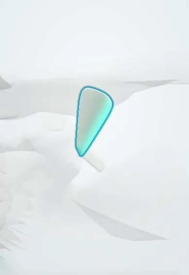
The conoid ligament ties the collarbone to the shoulder blade's coracoid process; it hangs the shoulder blade and stops the collarbone riding up.
Opens in the Durgical app, no sign-up needed.
What it is
The inner, cone-shaped coracoclavicular ligament from the coracoid to the underside of the collarbone; torn in higher-grade shoulder separations.
What it does
Hangs the shoulder blade from the collarbone and stops the collarbone riding up.
Key facts
- Attaches
- Base of the coracoid to the conoid tubercle of the clavicle
- Function
- Vertical AC stability; prevents upward displacement of the clavicle
- Tight in
- Arm elevation (posterior rotation of the clavicle)
Relations
- Above
- Conoid tubercle on the underside of the clavicle, near the junction of its lateral and middle thirds
- Below
- Base of the coracoid process, close to the suprascapular notch
- Lateral
- Trapezoid ligament, separated by fat and sometimes a small bursa
- Medial
- Subclavius under the clavicle; the suprascapular nerve passes nearby at the notch
How to find it
Not palpable. It rises almost vertically from the base of the coracoid, which lies deep below the clavicle in the infraclavicular hollow, to the conoid tubercle on the back of the underside of the clavicle near the junction of its lateral and middle thirds. Open repairs reach it by splitting the deltotrapezial fascia over the clavicle.
Examination
- Piano-key sign: in a complete AC separation the raised end of the clavicle can be pressed down and springs back up when released
On imaging
Bilateral AP or Zanca views of both AC joints compare the coracoclavicular distance; an axillary view checks for backward displacement of the clavicle (type IV).
Common problems
- Shoulder separation: a fall on the point of the shoulder sprains or tears the AC ligament, then the coracoclavicular ligaments.
- In severe cases the collarbone end sticks up as a visible bump.
- Later wear and arthritis of the AC joint.
How it is diagnosed
Tenderness and a step at the top of the shoulder. X-rays (sometimes comparing both sides) grade the separation.
Treatment
- Sling, ice and early motion for mild and moderate separations.
- Surgery to reconstruct the coracoclavicular ligaments for severe separations or ongoing pain.
Clinical notes
- Anatomic CC reconstruction rebuilds the conoid and the trapezoid with a tendon graft or suture loops passed under the coracoid and through two clavicle tunnels placed at their footprints
- Drilling or passing devices at the base of the coracoid puts the suprascapular nerve at risk, and the brachial plexus and axillary artery lie just medial and below
Anatomical variants
- A bony bridge or small joint can link the coracoid to the clavicle at the conoid area; one large study found it in roughly 1 in 11 people
Related conditions
Test yourself
Think of the answer, then tap the question to see it.
Which coracoclavicular ligament is the conoid ligament, and what does it mainly resist?
The posteromedial one; it mainly resists upward displacement of the clavicle.
On X-ray, which finding means the conoid and trapezoid ligaments are torn?
A coracoclavicular distance increased by more than about 25–50% compared with the other side.
What is the piano-key sign?
In a complete AC separation the raised clavicle end can be pressed down and springs back up.
Educational information, not medical advice. For any symptom, see a health professional.
Its names
- Official Latin (TA2)
- ligamentum conoideum TA2 1749
- Official Spanish (DeCS)
- Ligamento conoideo
- English (TA2)
- Conoid ligament
- Also called
- coracoclavicular ligament (conoid part)
Related
Sources: Terminologia Anatomica (TA2); Anatomy, Shoulder and Upper Limb, Acromioclavicular Joint; Anatomy of the Human Body (20th ed.): Surface Anatomy of the Upper Extremity (Lea & Febiger (Gray's Anatomy, 1918), 1918) · adapted, Public domain.
Spotted a mistake?We are always improving for the community. Tell us what is wrong and our team will review it.Report a mistake
Representative model, for educational purposes only.