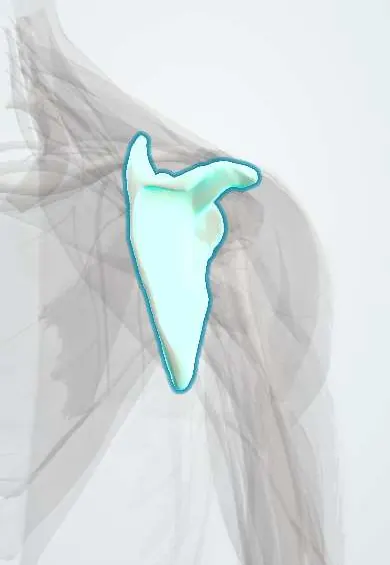
Scapula !
The scapula, or shoulder blade, is the flat bone on the upper back; it forms the shoulder socket and anchors the rotator cuff.
Opens in the Durgical app, no sign-up needed.
What it is
The flat triangular shoulder blade on the back of the ribs, with the acromion, coracoid and glenoid socket.
What it does
Gives the arm its socket and anchors the rotator cuff; it glides on the ribs as the arm rises.
Key facts
- Joints with
- Glenohumeral joint (humeral head); acromioclavicular joint (clavicle); scapulothoracic gliding plane on ribs 2–7
- Landmarks
- Spine, acromion, coracoid process, glenoid cavity; supra- and infraspinous fossae; superior angle T2, inferior angle T7; subscapular fossa; suprascapular notch; supraglenoid tubercle (long head of biceps) and infraglenoid tubercle (long head of triceps)
Relations
- In front
- Subscapularis fills the subscapular fossa; the serratus anterior inserts along the front of the medial border; both lie between the bone and the chest wall
- Behind
- Supraspinatus and infraspinatus in their fossae; the spine gives attachment to the middle trapezius and lower trapezius above and the rear deltoid below
- Medial
- Levator scapulae, rhomboid minor and rhomboid major on the medial border; the dorsal scapular nerve and artery run along it
- Lateral
- Long head of the triceps from the infraglenoid tubercle; teres minor and teres major from the lateral border, framing the quadrangular space (axillary nerve) and the triangular space (circumflex scapular artery)
- Above
- Coracoid process with the pectoralis minor, the short head of the biceps and the coracobrachialis; the suprascapular notch medial to its base
How to find it
The spine is followed from the acromion medially to its root at about the level of T3, and the inferior angle and medial border are felt with the arm relaxed. The coracoid process is felt by deep pressure just below the lateral third of the clavicle, in the deltopectoral groove; the acromion is the flat bony edge at the top of the shoulder.
Examination
- Wall push-up: the medial border and inferior angle lift away from the chest wall in serratus anterior weakness (long thoracic nerve)
- Resisted shoulder shrug and arm abduction: weakness with lateral winging and a drooping shoulder suggests spinal accessory nerve injury
On imaging
Shoulder trauma series: true AP of the glenohumeral joint, scapular Y view and axillary view. On the Y view the humeral head should sit over the centre of the Y (the glenoid); it lies in front in anterior dislocation and behind in posterior dislocation. CT with 3D reconstruction for glenoid and body fractures.
Common problems
- Fractures are uncommon and usually come from high-energy trauma.
- Poor control of the shoulder blade (scapular dyskinesis) can strain the shoulder.
- Winging when the serratus anterior or trapezius nerve is injured.
How it is diagnosed
Exam of how the shoulder blade moves as the arm rises. X-rays and CT for fractures.
Treatment
- Physical therapy to retrain the shoulder blade muscles.
- Surgery only for fractures that involve the socket or are badly displaced.
Clinical notes
- Suprascapular nerve compression at the suprascapular notch weakens supraspinatus and infraspinatus; at the spinoglenoid notch (often by a cyst from a labral tear) only infraspinatus is affected
- Bony Bankart lesion: a fracture of the anterior-inferior glenoid rim after anterior dislocation; glenoid bone loss raises the risk of recurrent dislocation
- The coracoid is a key surgical landmark: the brachial plexus and axillary vessels lie medial and deep to it
Anatomical variants
- Os acromiale: an acromial ossification centre that never fused, often on both sides; it can mimic a fracture and is linked with impingement
- Acromion shape varies (flat, curved, hooked); a hooked acromion is associated with subacromial impingement and rotator cuff tears
Development
The body ossifies before birth; the coracoid, the acromion (from several centres), the glenoid rim, the medial border and the inferior angle have secondary centres that fuse between late adolescence and the mid-twenties and can mimic fractures.
Memory aid
At the suprascapular notch: "the Army goes over the bridge, the Navy goes under" (artery over the ligament, nerve under it).
Related conditions
Test yourself
Think of the answer, then tap the question to see it.
At the suprascapular notch of the scapula, which structure passes under the suprascapular ligament and which passes above it?
The suprascapular nerve passes under the ligament and the suprascapular artery above it.
Medial winging of the scapula points to injury of which nerve and muscle?
The long thoracic nerve and the serratus anterior: the medial border and inferior angle lift off the chest, as on a wall push-up.
Lateral winging of the scapula with a drooping shoulder suggests injury of which nerve?
The spinal accessory nerve, which supplies the trapezius.
A scapular fracture should prompt a search for which associated injuries?
Rib fractures, pneumothorax, lung contusion and brachial plexus injury, because a scapular fracture marks high-energy trauma.
On the scapular Y view, where does the humeral head lie in anterior shoulder dislocation?
In front of the centre of the Y (the glenoid); in posterior dislocation it lies behind it.
Educational information, not medical advice. For any symptom, see a health professional.
Its names
- Official Latin (TA2)
- scapula TA2 1143
- Official Spanish (DeCS)
- Escápula
- English (TA2)
- Scapula
- Also called
- shoulder blade
Related
- AC capsule
- AC disc
- AC ligament
- Long biceps tendon
- Glenoid cartilage
- Humeral head cartilage
- Conoid ligament
- Coracoacromial ligament
Sources: Terminologia Anatomica (TA2); Anatomy, Thorax, Scapula.
Spotted a mistake?We are always improving for the community. Tell us what is wrong and our team will review it.Report a mistake
Representative model, for educational purposes only.