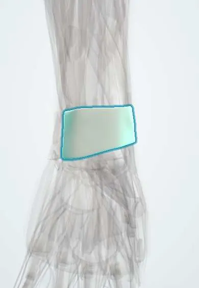
Extensor retinaculum !
The extensor retinaculum is a band across the back of the wrist that keeps the extensor tendons in six tunnels so they don't bowstring.
Official name: Extensor retinaculum of the wrist
Opens in the Durgical app, no sign-up needed.
What it is
A band across the back of the wrist, divided into six tunnels for the extensor tendons.
What it does
Keeps the extensor tendons from bowstringing when the wrist bends back.
Key facts
- Attaches
- Lateral distal radius to triquetrum, pisiform and ulnar styloid region
- Function
- Holds the extensor tendons in six compartments
- Tight in
- Wrist extension
Relations
- Contents
- Radial to ulnar: I APL and EPB; II ECRL and ECRB; III EPL; IV extensor digitorum and extensor indicis, with the posterior interosseous nerve; V extensor digiti minimi, over the DRUJ; VI ECU, in the groove of the ulna
- Superficial
- Branches of the superficial radial nerve and the dorsal branch of the ulnar nerve, and the back-of-hand veins
- Deep
- Dorsal distal radius, ulnar head and carpus; septa from its deep surface attach to the radius and separate the compartments
How to find it
Lister's tubercle is the bony bump on the back of the distal radius, roughly in line with the cleft between index and middle fingers. With the thumb extended, the anatomical snuffbox shows the tendons of the first (radial border) and third (ulnar border) compartments; the ECU tendon is felt in its groove beside the ulnar head with the wrist ulnarly deviated.
Examination
- Finkelstein test: the thumb is held in the palm and the wrist is deviated ulnarly; sharp pain over the radial styloid suggests de Quervain tenosynovitis of the first compartment
- ECU synergy test: resisted thumb abduction with the forearm supinated while ECU is palpated; pain along ECU suggests ECU tendinitis rather than a joint problem
- EPL lift test: with the palm flat, the thumb is lifted off the table; inability suggests EPL rupture
On imaging
Ultrasound identifies each compartment in short axis, using Lister's tubercle as the landmark between the second and third; it shows tenosynovitis, a septum dividing the first compartment, and ECU subluxation on dynamic supination.
Clinical notes
- De Quervain release: the first compartment is opened on its dorsal side, protecting branches of the superficial radial nerve; a missed separate EPB tunnel is a cause of failure
- Intersection syndrome: pain and crepitus a few centimetres proximal to the retinaculum, where the first-compartment muscles cross over the second-compartment tendons
- EPL rupture at Lister's tubercle after distal radius fractures and in rheumatoid arthritis
- ECU subluxation from a tear of its sixth-compartment subsheath, with a painful snap in supination and ulnar deviation
Anatomical variants
- A septum often divides the first compartment into separate tunnels for APL and EPB
- APL commonly has several tendon slips
- Extensor digitorum brevis manus, an extra muscle in the fourth compartment, can mimic a dorsal ganglion
Memory aid
Tendons per compartment, radial to ulnar: 2-2-1-2-1-1 («22-12-11»).
Related conditions
Test yourself
Think of the answer, then tap the question to see it.
How many compartments does the extensor retinaculum form, and which landmark separates the 2nd from the 3rd?
Six compartments; Lister's tubercle separates the 2nd from the 3rd.
Why does the extensor retinaculum not tether forearm rotation?
On the ulnar side it does not attach to the ulna but sweeps around to the pisiform and triquetrum.
What is the Finkelstein test?
The thumb is held in the palm and the wrist deviated ulnarly; sharp pain over the radial styloid suggests de Quervain tenosynovitis of the 1st compartment.
What is intersection syndrome?
Pain and crepitus a few centimetres proximal to the extensor retinaculum, where the 1st-compartment muscles cross over the 2nd-compartment tendons.
Educational information, not medical advice. For any symptom, see a health professional.
Its names
- Official Latin (TA2)
- retinaculum extensorium carpi TA2 2546
- Official Spanish (DeCS)
- Retináculo de los músculos extensores
- English (TA2)
- Extensor retinaculum of the wrist
- Also called
- dorsal carpal ligament
Related
Sources: Terminologia Anatomica (TA2); Anatomy, Shoulder and Upper Limb, Wrist Extensor Muscles.
Spotted a mistake?We are always improving for the community. Tell us what is wrong and our team will review it.Report a mistake
Representative model, for educational purposes only.