Cardiovascular system · Upper limb
Interosseous arteries !
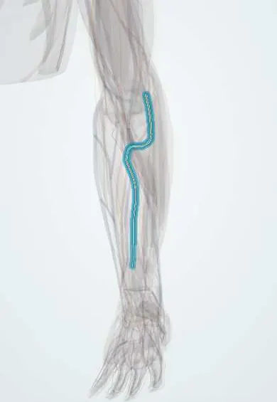
The interosseous arteries run along the membrane between the radius and ulna; they feed the deep muscles of the forearm.
Official name: Interosseous arteries of the forearm
Opens in the Durgical app, no sign-up needed.
What it is
Branches of the ulnar artery on both sides of the interosseous membrane.
What it does
Feed the deep forearm muscles.
Key facts
- Origin
- Common interosseous from the ulnar artery
- Branches
- Anterior and posterior interosseous, recurrent interosseous
- Supplies
- Deep flexors and extensors of the forearm, radius and ulna
Relations
- Course
- Short common trunk; the anterior artery descends on the membrane to pronator quadratus; the posterior artery passes above the membrane, between it and the oblique cord, to the back of the forearm and gives the recurrent interosseous artery up to the elbow
- In front
- Over the anterior artery: flexor digitorum profundus (FDP) and flexor pollicis longus (FPL), with the anterior interosseous nerve beside it
- Behind
- Around the posterior artery: the superficial extensors (extensor digitorum, ECU) behind it and the deep group (APL, EPB, EPL, extensor indicis) in front of it
- Deep
- The interosseous membrane, between the two arteries
How to find it
Not palpable. The common trunk leaves the ulnar artery just below the radial tuberosity. The anterior artery lies on the front of the interosseous membrane with its nerve; the posterior artery appears between supinator and abductor pollicis longus and runs between the superficial and deep extensors along a line from the lateral epicondyle to the distal radioulnar joint, the axis of its flap.
On imaging
Not imaged on their own in routine practice. On ultrasound or angiography the common interosseous artery leaves the ulnar artery just below the elbow and divides into anterior and posterior branches on either side of the interosseous membrane; the anterior one runs with the anterior interosseous nerve. Angiography shows them carrying collateral flow when the radial or ulnar artery is blocked.
Clinical notes
- In forearm compartment syndrome and Volkmann contracture, the deep flexors fed by the anterior artery (FDP and FPL) are the most affected
- Reverse posterior interosseous artery flap: skin from the back of the forearm turned down on this artery to cover defects on the back of the hand
- Their end branches join the dorsal carpal network, a collateral route to the wrist when the radial or ulnar artery is blocked
Anatomical variants
- Persistent median artery: a branch of the anterior interosseous artery that runs with the median nerve and sometimes reaches the palm through the carpal tunnel
Development
The anterior interosseous artery is a remnant of the embryonic axis artery of the forearm; the median artery that arises from it normally regresses before birth.
Test yourself
Think of the answer, then tap the question to see it.
Where does the common interosseous artery arise, and how does it divide?
From the ulnar artery just below the radial tuberosity; it splits into anterior and posterior interosseous arteries at the upper border of the interosseous membrane.
Which nerve accompanies each interosseous artery?
The anterior interosseous artery runs with the anterior interosseous nerve; the posterior with the posterior interosseous nerve.
Which muscles fed by the anterior interosseous artery are most affected in forearm compartment syndrome?
The deep flexors: flexor digitorum profundus and flexor pollicis longus.
Educational information, not medical advice. For any symptom, see a health professional.
Its names
- Official Latin (TA2)
- arteria interossea communis TA2 4661
- Official Spanish (DeCS)
- Arterias interóseas común, anterior, posterior y recurrente
- English (TA2)
- Interosseous arteries of the forearm
Related
Sources: Terminologia Anatomica (TA2); Anatomy, Shoulder and Upper Limb, Ulnar Artery; Anatomy, Shoulder and Upper Limb, Forearm Arteries; Anatomy of the Human Body (20th ed.): Development of the Vascular System (Lea & Febiger (Gray's Anatomy, 1918), 1918) · adapted, Public domain.
Spotted a mistake?We are always improving for the community. Tell us what is wrong and our team will review it.Report a mistake
Representative model, for educational purposes only.