Cardiovascular system · Lower limb
External iliac vein !
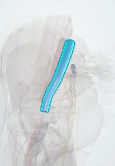
The external iliac vein carries blood from the leg up into the pelvis; it continues from the femoral vein at the groin.
Opens in the Durgical app, no sign-up needed.
What it is
A large vein inside the pelvis, continuing from the femoral vein above the groin.
What it does
Returns blood from the leg toward the heart.
Key facts
- Drains
- Lower limb via femoral vein; inferior epigastric and deep circumflex iliac veins
- Into
- Common iliac vein, joining internal iliac vein in front of sacroiliac joint
Relations
- Course
- Continues from the femoral vein behind the inguinal ligament, runs up along the pelvic brim and unites with the internal iliac vein in front of the sacroiliac joint, forming the common iliac vein
- Lateral
- The external iliac artery at the groin; higher up the vein passes more behind the artery
- Medial
- External iliac lymph nodes and the pelvic side wall, along which the obturator nerve runs below
- In front
- Crossed by the ureter near the pelvic brim and by the ductus deferens (or round ligament) near the deep inguinal ring
How to find it
Not palpable. It follows the pelvic brim on the edge of the psoas, partly behind its artery at first and fully on its medial side lower down. In pelvic lymph node dissection it is exposed beneath the external iliac nodes; on ultrasound it is traced upward from the common femoral vein above the inguinal ligament, though bowel gas often hides it.
On imaging
Bowel gas limits ultrasound in the pelvis. A common femoral vein Doppler signal that loses its normal variation with breathing suggests a blockage higher up, in the iliac veins. CT or MR venography shows the vein directly.
Common problems
- A deep vein clot (DVT) in the leg can extend up into it.
Clinical notes
- Kidney transplant: the donor renal vein is usually joined to it
- Chronic iliac vein obstruction, after DVT or from compression, can be reopened with venous stents
- Injured in pelvic fractures and in pelvic lymph node surgery, where it lies beneath the nodes
Related conditions
Test yourself
Think of the answer, then tap the question to see it.
Why is left-sided iliofemoral DVT more common?
The right common iliac artery crosses and can compress the left common iliac vein (May–Thurner).
Which Doppler finding in the common femoral vein suggests a blockage of the iliac veins?
Loss of the normal variation of the signal with breathing.
Where does the external iliac vein end?
In front of the sacroiliac joint it unites with the internal iliac vein, and together they become the common iliac vein.
Educational information, not medical advice. For any symptom, see a health professional.
Its names
- Official Latin (TA2)
- vena iliaca externa TA2 5050
- Official Spanish (DeCS)
- Vena ilíaca externa DeCS D007084
- English (TA2)
- External iliac vein
- Also called
- vena ilica externa
Related
Sources: Terminologia Anatomica (TA2); DeCS; Anatomy, Abdomen and Pelvis: Veins; Anatomy of the Human Body (20th ed.): The External Iliac Artery (Lea & Febiger (Gray's Anatomy, 1918), 1918) · adapted, Public domain.
Spotted a mistake?We are always improving for the community. Tell us what is wrong and our team will review it.Report a mistake
Representative model, for educational purposes only.