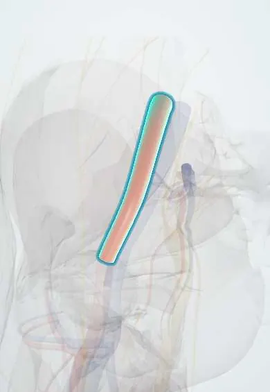
Cardiovascular system · Lower limb
External iliac artery !
The external iliac artery is the main artery to the leg inside the pelvis; it becomes the femoral artery at the groin.
Opens in the Durgical app, no sign-up needed.
What it is
A large artery inside the pelvis, branching from the common iliac artery and running along the psoas muscle.
What it does
Carries blood to the leg. It becomes the femoral artery as it passes under the inguinal ligament.
Key facts
- Origin
- Common iliac artery at level of sacroiliac joint (pelvic brim)
- Branches
- Inferior epigastric, deep circumflex iliac
- Supplies
- Lower limb; lower anterior abdominal wall via branches; becomes femoral behind inguinal ligament
Relations
- Course
- From the common iliac bifurcation in front of the sacroiliac joint it runs down and forward along the pelvic brim and passes behind the inguinal ligament at the mid-inguinal point to become the femoral artery
- Lateral
- The psoas major, with the genitofemoral nerve on it
- Medial
- The external iliac vein, medial to it at the inguinal ligament and partly behind it higher up; the external iliac lymph nodes around both
- In front
- Peritoneum; crossed by the ureter near its origin, by the gonadal vessels and, near the deep ring, by the ductus deferens (or round ligament); the ileum on the right, the sigmoid colon on the left
How to find it
Not felt. Its surface line runs from about 2 cm below and slightly left of the umbilicus (the aortic bifurcation, about L4) to the mid-inguinal point: the upper third of this line marks the common iliac artery and the lower two-thirds the external iliac artery.
On imaging
CT angiography is the standard to map stenosis, occlusion or aneurysm from the iliac bifurcation to the inguinal ligament; MR angiography is an alternative. Duplex ultrasound sees it through the abdomen and shows stenosis as high velocity. It is a key access route for endovascular repair, so its calibre and calcium are checked on CT first. Radiographs only show calcified walls.
Common problems
- Narrowing from plaque (peripheral artery disease) can cause leg or buttock pain when walking.
Clinical notes
- A femoral puncture made too high enters the external iliac artery: bleeding then tracks into the retroperitoneum, cannot be compressed and may show only as falling blood pressure or flank and back pain
- Aortoiliac occlusive disease (Leriche syndrome): buttock and thigh claudication, weak or absent femoral pulses and erectile dysfunction
- Route for iliac angioplasty and stents and for large-bore delivery systems such as those for aortic stent grafts and heart valves
- Its deep circumflex iliac branch runs behind the inguinal ligament to the anterior superior iliac spine and then along the inner lip of the iliac crest; it is the pedicle of the vascularised iliac crest flap, and its ascending branch can bleed at lateral laparoscopic ports
Anatomical variants
- Agenesis (rare): the internal iliac artery then continues into the thigh and takes over the supply of the leg
- Marked coiling or tortuosity (rare), which matters in catheter procedures
- Its inferior epigastric branch may give rise to the obturator artery (an aberrant obturator artery)
Development
The early lower limb bud is fed by an axial artery that runs with the sciatic nerve and comes from the internal iliac side. The external iliac and femoral arteries grow later and take over; when the axial vessel survives into adult life it is called a persistent sciatic artery.
Related conditions
Test yourself
Think of the answer, then tap the question to see it.
Where does the external iliac artery become the femoral artery?
Behind the inguinal ligament, at the mid-inguinal point.
Which branch of the external iliac artery separates indirect from direct inguinal hernias?
The inferior epigastric artery: indirect hernias pass lateral to it, direct hernias medial to it through Hesselbach's triangle.
Why is a femoral puncture made too high dangerous?
It enters the external iliac artery: bleeding tracks into the retroperitoneum, cannot be compressed and may show only as falling blood pressure or flank pain.
Which artery is the donor renal artery usually joined to in a kidney transplant?
The external iliac artery, with the kidney placed in the iliac fossa.
Educational information, not medical advice. For any symptom, see a health professional.
Its names
- Official Latin (TA2)
- arteria iliaca externa TA2 4357
- Official Spanish (DeCS)
- Arteria ilíaca externa DeCS D007083
- English (TA2)
- External iliac artery
- Also called
- arteria ilica externa
Related
Sources: Terminologia Anatomica (TA2); DeCS; Anatomy, Abdomen and Pelvis: External Iliac Arteries; Tratado de anatomía humana (9.ª ed.), tomo II, Angiología: aorta descendente, arterias ilíacas y arterias del miembro inferior.
Spotted a mistake?We are always improving for the community. Tell us what is wrong and our team will review it.Report a mistake
Representative model, for educational purposes only.