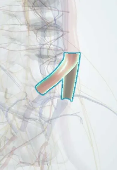
Common iliac arteries !
The common iliac arteries are the two branches of the aorta, starting at about L4; they carry blood to the pelvis and legs.
Official name: Common iliac artery
Opens in the Durgical app, no sign-up needed.
What it is
The two branches of the aorta, starting at about L4 and running down the sides of the pelvis.
What it does
Carry blood to the pelvis and legs. The L5-S1 disc is usually reached between them in ALIF.
Key facts
- Origin
- Bifurcation of the abdominal aorta at L4, slightly left of the midline
- Branches
- Internal and external iliac arteries, dividing in front of the sacroiliac joint (L5–S1); small twigs to psoas and ureter
- Supplies
- Pelvis, perineum, gluteal region and lower limbs
Relations
- Course
- From the aortic bifurcation at L4 each runs down and out along the medial border of the psoas major and ends in front of the sacroiliac joint by dividing into internal and external iliac arteries
- In front
- Peritoneum and small bowel, the ureter near its end and, on the left, the sigmoid mesocolon and superior rectal vessels
- Behind
- L4 and L5 vertebral bodies and the L4–L5 disc, the sympathetic trunk and the lumbosacral trunk; the left common iliac vein and the start of the inferior vena cava lie behind the right artery
- Lateral
- Psoas major
- Medial
- Superior hypogastric plexus and the median sacral vessels in the gap between the two arteries; the left common iliac vein lies medial to the left artery
How to find it
On the surface, a line from just below and left of the umbilicus (the bifurcation) to the mid-inguinal point, halfway from the anterior superior iliac spine to the pubic symphysis, marks the iliac arteries; its upper third is the common iliac artery.
On imaging
CT angiography is the main test: it shows aneurysms, often with an aortic one, and narrowing or blockage in aortoiliac occlusive disease; Doppler ultrasound shows flow, and radiographs may show wall calcification. On CT or MR venography, the right common iliac artery is seen pressing the left common iliac vein against L5 (May–Thurner); intravascular ultrasound confirms it.
Clinical notes
- Long retraction of the left common iliac artery in anterior L4–L5 surgery can cause thrombosis and a cold, pulseless leg, so foot pulses or toe oxygen saturation are watched during and after the operation
- The plexus fibres in front of L5 are swept aside bluntly, with little or no monopolar cautery, to lower the risk of retrograde ejaculation
- Atherosclerotic narrowing (aortoiliac disease) causes buttock and thigh claudication and weak femoral pulses; common iliac aneurysms also occur, often together with an aortic aneurysm
Anatomical variants
- Their origin follows the bifurcation, usually at L4 but ranging from about L3 to L5; a high or low bifurcation changes the size of the window to the L5–S1 disc
Related conditions
Test yourself
Think of the answer, then tap the question to see it.
Where do the common iliac arteries begin and end?
They begin at the aortic bifurcation at L4 and end in front of the sacroiliac joint by dividing into internal and external iliac arteries.
Which artery compresses the left common iliac vein in May-Thurner syndrome?
The right common iliac artery, which crosses in front of the left common iliac vein over L5; it can lead to left leg deep vein thrombosis.
Which structure crosses the end of the common iliac artery at the pelvic brim?
The ureter: a landmark in pelvic and anterior spine surgery.
Which complication follows damage to the superior hypogastric plexus during anterior L5–S1 exposure?
Retrograde ejaculation; the plexus lies in front of L5 between the two common iliac arteries.
Educational information, not medical advice. For any symptom, see a health professional.
Its names
- Official Latin (TA2)
- arteria iliaca communis TA2 4301
- Official Spanish (DeCS)
- Arterias ilíacas comunes DeCS D007083
- English (TA2)
- Common iliac artery
- Also called
- arteria ilica communis
Related
Sources: Terminologia Anatomica (TA2); DeCS; Anatomy, Abdomen and Pelvis: Arteries; Tratado de anatomía humana (9.ª ed.), tomo II, Angiología: aorta descendente, arterias ilíacas y arterias del miembro inferior.
Spotted a mistake?We are always improving for the community. Tell us what is wrong and our team will review it.Report a mistake
Representative model, for educational purposes only.