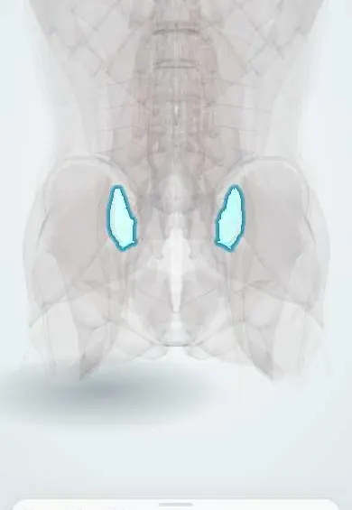
SI joint surfaces !
The sacroiliac joint surfaces are ear-shaped, cartilage-covered areas between the sacrum and hip bones that pass the spine's weight to the pelvis.
Official name: Sacroiliac joint
Opens in the Durgical app, no sign-up needed.
What it is
The cartilage-covered, ear-shaped joint surfaces between the sacrum and the hip bones.
What it does
Where the weight of the spine passes into the pelvis; the joint moves only a few degrees.
Key facts
- Where
- Ear-shaped (auricular) surfaces of the sacrum (S1–S3) and the ilium
- Attaches
- Sacral side: thicker hyaline cartilage; iliac side: thinner, more fibrous cartilage; held by anterior, interosseous and posterior sacroiliac ligaments
- Function
- Synovial joint in front, fibrous (syndesmosis) behind; passes body weight from spine to pelvis; only a few degrees of nutation and counternutation
Relations
- Medial
- Auricular surface of the sacrum (S1–S3), with the sacral foramina and the S1–S2 roots just medial
- Lateral
- Auricular surface of the ilium, with the iliac tuberosity behind it for the interosseous ligament
- In front
- Anterior sacroiliac ligament; in front, psoas major, the lumbosacral trunk and the obturator nerve crossing the pelvic brim, and the internal iliac vessels
- Behind
- Interosseous and posterior sacroiliac ligaments, then the thoracolumbar fascia and gluteus maximus
How to find it
The joint is deep and faces forward and outward, so it cannot be felt directly. The posterior superior iliac spines are felt under the skin dimples above the buttocks; the joint lies deep and a little below and medial to each one.
Examination
- FABER (Patrick) test: supine, the hip is flexed, abducted and externally rotated (figure 4) and the knee pressed down; pain at the back over the SI region suggests the SI joint, groin pain suggests the hip
- Gaenslen test: one hip is held flexed to the chest while the other leg hangs extended over the edge of the table; pain in the SI region suggests the SI joint
- Thigh thrust, distraction, compression and sacral thrust complete the Laslett cluster: three or more positive tests out of five raise the probability that the SI joint is the pain source
- Fortin finger test: the person points with one finger to the pain within about 1 cm below and medial to the PSIS; pointing there consistently suggests the SI joint
On imaging
AP pelvis or angled (Ferguson) view: erosions, sclerosis, joint widening and later fusion. MRI (STIR or fat-suppressed T2) shows bone marrow oedema of active sacroiliitis before radiographs change. CT shows erosions and sacral insufficiency fractures in older people.
Common problems
- SI joint pain: low back and buttock pain, sometimes into the thigh, from strain, a fall, uneven loading, pregnancy or after a lumbar fusion.
- Inflammation of the joint (sacroiliitis), for example in ankylosing spondylitis.
How it is diagnosed
Exam with pain provocation tests; a numbing injection into the joint confirms it. X-rays, CT or MRI for arthritis and inflammation.
Treatment
- Physical therapy, a pelvic belt, medicine and joint injections.
- Radiofrequency ablation of the nerves, or SI joint fusion surgery for lasting severe pain.
Clinical notes
- Bilateral, symmetric sacroiliitis is typical of ankylosing spondylitis and inflammatory bowel disease; it is often asymmetric in psoriatic and reactive arthritis
- Septic sacroiliitis is usually one-sided and can mimic disc or hip disease
- Hormonal laxity in pregnancy contributes to pelvic girdle pain
- An intra-articular block with local anaesthetic is the reference test for SI joint pain
Anatomical variants
- Accessory sacroiliac joints, small extra contacts between ilium and sacrum outside the main joint, are common (reported in about 8–40% of specimens)
Development
In children the surfaces are flat and mobile; from adolescence they develop interlocking ridges and hollows, and fibrous bridging becomes common in older age.
Related conditions
Test yourself
Think of the answer, then tap the question to see it.
On which side of the sacroiliac joint surfaces do the erosions of sacroiliitis start, and why?
On the iliac side, where the cartilage is thinner.
How is the FABER (Patrick) test done, and how is it read for the sacroiliac joint?
Supine, the hip is flexed, abducted and externally rotated and the knee pressed down; pain at the back over the SI region suggests the SI joint, groin pain the hip.
What is the Laslett cluster for sacroiliac joint pain?
Five provocation tests (distraction, compression, thigh thrust, sacral thrust, Gaenslen); three or more positive raise the probability that the SI joint is the source.
What is the reference test for sacroiliac joint pain?
An intra-articular block with local anaesthetic.
Educational information, not medical advice. For any symptom, see a health professional.
Its names
- Official Latin (TA2)
- articulatio sacroiliaca TA2 1861
- Official Spanish (DeCS)
- Articulación sacroiliaca DeCS D012446
- English (TA2)
- Sacroiliac joint
- Also called
- articulus sacrioilicus
Related
Sources: Terminologia Anatomica (TA2); DeCS; Anatomy, Abdomen and Pelvis, Sacroiliac Joint.
Spotted a mistake?We are always improving for the community. Tell us what is wrong and our team will review it.Report a mistake
Representative model, for educational purposes only.