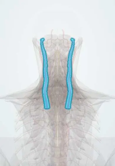
Internal jugular vein !
The internal jugular vein is the large neck vein beside the carotid artery; it drains blood from the brain, face and neck.
Opens in the Durgical app, no sign-up needed.
What it is
The large vein in the neck beside the carotid artery, inside the carotid sheath.
What it does
Drains blood from the brain, face and neck.
Key facts
- Drains
- Brain (dural venous sinuses), face and neck; begins at the jugular foramen as continuation of the sigmoid sinus
- Into
- Joins the subclavian vein behind the sternal end of the clavicle to form the brachiocephalic vein
Relations
- Course
- From the jugular foramen it runs down inside the carotid sheath, lateral to the internal and then the common carotid artery and under the sternocleidomastoid, to the root of the neck
- Medial
- Internal and then common carotid artery, with the vagus nerve behind, between them
- Behind
- Cervical transverse processes and the sympathetic trunk; lower down the scalenus anterior with the phrenic nerve, the first part of the subclavian artery and the dome of the pleura
- Superficial
- Sternocleidomastoid, the omohyoid crossing it, and the deep cervical lymph nodes along its length
How to find it
It lies under the sternocleidomastoid; its lower end sits in the small triangle between the sternal and clavicular heads, just above the clavicle and lateral to the carotid pulse. The venous pulse is looked for with the trunk at about 45° and the head turned slightly away.
Examination
- Jugular venous pressure: the vertical height of the top of the venous pulsation above the sternal angle; more than about 3–4 cm suggests raised right heart filling pressure
- Abdominojugular (hepatojugular) reflux: firm, sustained pressure on the abdomen gives a sustained rise of the venous pulse in right heart failure
- Kussmaul sign: the venous pressure rises instead of falling on breathing in, as in constrictive pericarditis
On imaging
On ultrasound it is larger than the carotid and lateral to it, collapses with light probe pressure and fills with a Valsalva manoeuvre; a clot makes it non-compressible.
Clinical notes
- Line complications include carotid puncture, pneumothorax with low punctures and air embolism; a head-down tilt while the line is placed lowers the air risk
- Lemierre syndrome: septic thrombophlebitis of the vein after a throat infection, often with septic emboli to the lungs
- Removed in radical neck dissection; removing both veins at once risks facial and brain swelling, so it is usually avoided
Anatomical variants
- The right vein is usually larger than the left
- One vein can be small or even absent, which matters when choosing a side for a central line
Development
It comes from the anterior cardinal (primitive jugular) vein; a cross channel between the two sides becomes the left brachiocephalic vein, so the right vein runs straight down into the superior vena cava.
Memory aid
Carotid sheath: "I See 10 CC's in the IV": Internal Carotid, cranial nerve 10 (vagus), Common Carotid, Internal jugular Vein.
Related conditions
Test yourself
Think of the answer, then tap the question to see it.
Where does the internal jugular vein begin and end?
It begins at the jugular foramen from the sigmoid sinus and joins the subclavian vein behind the sternal end of the clavicle to form the brachiocephalic vein.
What is the classic landmark for internal jugular vein cannulation?
The apex of the triangle between the two heads of the sternocleidomastoid and the clavicle, lateral to the carotid pulse; ultrasound guidance is now standard.
Why is the right internal jugular vein used to read the jugular venous pressure?
It runs in a straight line down to the superior vena cava and right atrium.
What is Lemierre syndrome?
Septic thrombophlebitis of the internal jugular vein after a throat infection, often with septic emboli to the lungs.
What is the Kussmaul sign in the internal jugular vein?
The venous pressure rises instead of falling on breathing in, as in constrictive pericarditis.
Educational information, not medical advice. For any symptom, see a health professional.
Its names
- Official Latin (TA2)
- vena jugularis interna TA2 4800
- Official Spanish (DeCS)
- Vena yugular interna DeCS D007601
- English (TA2)
- Internal jugular vein
Related
Sources: Terminologia Anatomica (TA2); DeCS; Anatomy, Head and Neck: Internal Jugular Vein; Anatomy of the Human Body (20th ed.): Development of the Vascular System (Lea & Febiger (Gray's Anatomy, 1918), 1918) · adapted, Public domain; Tratado de anatomía humana (9.ª ed.), tomo II, Libro IV Angiología, sección 4: Venas (sistemas de las venas cavas superior e inferior, vena porta, venas del raquis).
Spotted a mistake?We are always improving for the community. Tell us what is wrong and our team will review it.Report a mistake
Representative model, for educational purposes only.