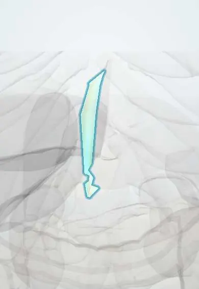
Falciform ligament !
The falciform ligament is a thin, sickle-shaped fold that attaches the front of the liver to the abdominal wall and separates its right and left lobes.
Opens in the Durgical app, no sign-up needed.
What it is
A thin fold of peritoneum attaching the liver to the front abdominal wall, carrying the round ligament (the remnant of the umbilical vein).
What it does
Divides the surface of the liver into right and left; in cirrhosis veins around it can widen.
Key facts
- Where
- Sickle-shaped peritoneal fold from anterior abdominal wall above umbilicus and diaphragm to anterosuperior liver surface
- Attaches
- Free edge holds round ligament of liver (obliterated umbilical vein) and paraumbilical veins; layers part as coronary and left triangular ligaments
- Function
- Divides anatomical right and left lobes (segments II–III from IV); paraumbilical veins form a portosystemic anastomosis
Relations
- In front
- Back of the anterior abdominal wall from the umbilicus up to the diaphragm, a little to the right of the midline
- Behind
- Anterior and upper surfaces of the liver, between liver segment IV on the right and segments II–III on the left
- Above
- Diaphragm; at the top its two layers part to form the coronary ligament (right) and the left triangular ligament, around the bare area
- Below
- Free lower edge, ending at the umbilical fissure where the round ligament enters the liver
How to find it
Runs from the umbilicus up to the front and upper surface of the liver, a little right of the midline, and divides the classical right and left lobes on the front of the liver. At laparoscopy it hangs from the anterior abdominal wall as a sickle-shaped fold; an upper midline incision passes right beside it.
Examination
- Flow direction in dilated abdominal wall veins: a vein is emptied between two fingers and one end released; flow away from the umbilicus fits portal hypertension, flow upward from below fits inferior vena cava obstruction
On imaging
With free air in the abdomen it can be outlined on a supine X-ray as a bright line (falciform ligament sign), part of the football sign in infants. On ultrasound and CT the fat around the round ligament marks the umbilical fissure between segments III and IV; Doppler shows a recanalized umbilical vein.
Common problems
- Fatty liver disease, very common with obesity and diabetes.
- Hepatitis (viral, alcohol).
- Cirrhosis: scarring that can lead to liver failure and portal hypertension.
- Liver cancer and metastases.
How it is diagnosed
Blood tests (liver enzymes), ultrasound, elastography, CT or MRI, biopsy.
Treatment
- Treat the cause (weight loss, antiviral medicines, stopping alcohol); surgery to remove segments; liver transplant for failure.
Clinical notes
- In newborns an umbilical venous catheter follows the fetal route: umbilical vein along the free edge of this ligament, left portal vein, ductus venosus, then the inferior vena cava
- In an upper midline laparotomy the ligament is usually divided between ties, because a reopened umbilical vein in its free edge can bleed briskly in portal hypertension
- Free air under the abdominal wall can outline the ligament on a supine radiograph (football sign), a clue to perforation, especially in newborns
Development
Comes from the ventral mesogastrium between the liver and the anterior abdominal wall; its free edge carries the left umbilical vein, which brings oxygenated blood from the placenta and closes after birth to become the round ligament.
Test yourself
Think of the answer, then tap the question to see it.
What runs in the free edge of the falciform ligament?
The round ligament of the liver (obliterated umbilical vein) with the paraumbilical veins.
How does the falciform ligament relate to caput medusae?
In portal hypertension the paraumbilical veins along it open up, linking the left portal vein to the wall veins around the umbilicus.
Does the falciform ligament divide the functional right and left liver?
No: it divides the anatomical lobes (segments II–III from IV); the functional division is Cantlie line.
Educational information, not medical advice. For any symptom, see a health professional.
Its names
- Official Latin (TA2)
- ligamentum falciforme; ligamentum teres hepatis TA2 3771
- Official Spanish (DeCS)
- Ligamento falciforme
- English (TA2)
- Falciform ligament
- Also called
- mesohepaticum ventrale
Sources: Terminologia Anatomica (TA2); Anatomy, Abdomen and Pelvis: Falciform Ligament; 23.6 Accessory Organs in Digestion: The Liver, Pancreas, and Gallbladder (OpenStax (Rice University), 2022) · adapted, CC BY 4.0.
Spotted a mistake?We are always improving for the community. Tell us what is wrong and our team will review it.Report a mistake
Representative model, for educational purposes only.