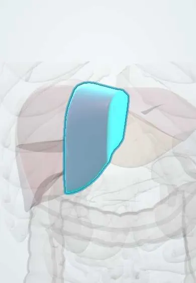
Liver segment IV !
Liver segment IV is the middle part of the left liver, between the falciform ligament and the gallbladder; its lower part forms part of the gallbladder bed.
Official name: Left medial segment of liver
Opens in the Durgical app, no sign-up needed.
What it is
Liver segment IV: the left medial part, including the quadrate lobe.
What it does
Because each segment has its own supply, surgeons can remove one or more segments and leave the rest working.
Key facts
- Where
- Left medial section between falciform ligament and Cantlie line (gallbladder–IVC); IVa superior, IVb inferior (quadrate lobe)
- Blood supply
- Branches of left portal vein (umbilical part) and left (or middle) hepatic artery; drains mainly into middle hepatic vein
- Nerve
- Hepatic plexus (celiac sympathetic and vagal fibres) along its portal pedicle, shared with the whole liver
- Function
- Functionally part of the left liver though it lies right of falciform ligament; bile to left hepatic duct
Relations
- Above
- IVa reaches up under the diaphragm to the confluence of the hepatic veins with the inferior vena cava
- Below
- IVb (quadrate lobe) sits on the porta hepatis, with the gallbladder fossa to its right, the umbilical fissure and round ligament to its left, and the pylorus and first part of the duodenum below
- Behind
- Liver segment I (caudate lobe), behind the porta hepatis
How to find it
Right of the falciform ligament, under the xiphoid process: IVb (quadrate lobe) is seen from below between the gallbladder and the round ligament, while IVa lies high under the diaphragm near the hepatic vein confluence.
On imaging
On CT or MRI it lies between the middle hepatic vein and the umbilical fissure, split into IVa above and IVb below the portal plane. Focal fatty sparing or fatty change near the falciform ligament and the gallbladder bed is a common pseudolesion here.
Common problems
- Fatty liver disease, very common with obesity and diabetes.
- Hepatitis (viral, alcohol).
- Cirrhosis: scarring that can lead to liver failure and portal hypertension.
- Liver cancer and metastases.
How it is diagnosed
Blood tests (liver enzymes), ultrasound, elastography, CT or MRI, biopsy.
Treatment
- Treat the cause (weight loss, antiviral medicines, stopping alcohol); surgery to remove segments; liver transplant for failure.
Clinical notes
- Its portal pedicles arise from the right side of the umbilical portion of the left portal vein, so a lesion in the umbilical fissure can threaten its blood supply
- In a classic split-liver graft it goes with the extended right graft, yet its inflow comes from the left side, so segment IV can lose blood supply after the split
Development
Its left border, the umbilical fissure, holds the round ligament, the closed fetal left umbilical vein; in portal hypertension small paraumbilical veins along it can reopen.
Related conditions
Test yourself
Think of the answer, then tap the question to see it.
Why is liver segment IV functionally left liver even though it lies right of the falciform ligament?
Its portal pedicles come from the umbilical part of the left portal vein and its bile drains to the left hepatic duct; Cantlie line is its right border.
Which liver segments are removed with the gallbladder bed in radical gallbladder cancer surgery?
Segment IVb and segment V, which form the gallbladder bed, with the regional nodes.
Which vein runs along the right border of liver segment IV?
The middle hepatic vein, in Cantlie plane; segment IV drains mainly into it.
Educational information, not medical advice. For any symptom, see a health professional.
Its names
- Official Latin (TA2)
- segmentum mediale sinistrum hepatis TA2 3071
- Official Spanish (DeCS)
- Segmento IV del hígado DeCS D008099
- English (TA2)
- Left medial segment of liver
- Also called
- segment IV of liver
Sources: Terminologia Anatomica (TA2); DeCS; Diagnostic Imaging of the Liver; Anatomy, Abdomen and Pelvis: Liver; 23.6 Accessory Organs in Digestion: The Liver, Pancreas, and Gallbladder (OpenStax (Rice University), 2022) · adapted, CC BY 4.0.
Spotted a mistake?We are always improving for the community. Tell us what is wrong and our team will review it.Report a mistake
Representative model, for educational purposes only.