Cardiovascular system · Abdomen
Portal vein !
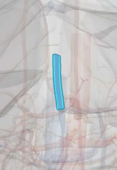
The portal vein carries blood from the intestines, stomach, pancreas and spleen into the liver, which processes the absorbed nutrients and toxins.
Official name: Hepatic portal vein
Opens in the Durgical app, no sign-up needed.
What it is
A large vein formed behind the pancreas by the splenic and superior mesenteric veins, running up to the liver.
What it does
Brings nutrient-rich blood from the stomach, intestines, pancreas and spleen to the liver, about three-quarters of the liver's blood supply.
Key facts
- Drains
- Formed behind pancreatic neck (L1–L2) by SMV and splenic vein; drains GI tract from lower esophagus to upper anal canal, spleen, pancreas, gallbladder
- Into
- Liver at porta hepatis, dividing into right and left branches; runs behind bile duct and hepatic artery in hepatoduodenal ligament
Relations
- Course
- From behind the neck of the pancreas it climbs up and to the right behind the first part of the duodenum, enters the hepatoduodenal ligament and reaches the porta hepatis, where it splits into right and left branches
- In front
- Neck of the pancreas and first part of the duodenum low down; higher up, the common bile duct and the proper hepatic artery in the ligament
- Behind
- Inferior vena cava, separated from it by the omental (epiploic) foramen, whose front wall is the free edge of the ligament carrying the portal vein
How to find it
Not palpable. At surgery it is found by feeling the free edge of the lesser omentum through the omental foramen: the vein is the deepest, softest structure of the triad. On ultrasound it is followed from the splenic–mesenteric confluence behind the pancreas up to the porta hepatis.
On imaging
Doppler ultrasound shows normal flow toward the liver (hepatopetal), continuous with mild respiratory variation; slow, to-and-fro or reversed (hepatofugal) flow points to portal hypertension, and an echogenic filling defect to thrombosis. Contrast CT or MRI in the portal venous phase maps the vein, its branches and any collaterals.
Common problems
- Portal hypertension in cirrhosis: blood backs up, causing swollen veins in the esophagus (varices) that can bleed, an enlarged spleen and fluid in the abdomen (ascites).
How it is diagnosed
Ultrasound with Doppler, CT, endoscopy for varices.
Treatment
- Medicines that lower the pressure, banding of varices, a shunt (TIPS), liver transplant.
Clinical notes
- Portal hypertension opens the anastomoses: left gastric to esophageal veins (esophageal varices), paraumbilical to abdominal wall veins (caput medusae), superior to middle and inferior rectal veins (rectal varices), and retroperitoneal veins of the colon and bare area of the liver to renal and lumbar veins (veins of Retzius)
- TIPS: a stented tract through the liver from a hepatic vein to a portal vein branch lowers portal pressure; it can worsen hepatic encephalopathy because blood bypasses the liver
- Portal vein thrombosis (cirrhosis, clotting disorders, pancreatitis, newborn umbilical catheters) can turn into a web of collaterals at the porta hepatis (cavernous transformation)
- Involvement of the portal vein or the confluence by a cancer of the head of the pancreas is a key factor in judging whether it can be removed
Anatomical variants
- Branching variants, such as a trifurcation into left, right anterior and right posterior branches or a right posterior branch arising on its own, in up to about 35%; they matter in liver resection and living donor transplant
- Rarely, a preduodenal portal vein running in front of the duodenum, usually with malrotation or other laterality anomalies
Development
Forms from the vitelline veins. Before birth the left umbilical vein joins its left branch and the ductus venosus carries blood on to the inferior vena cava; they become the ligamentum teres and the ligamentum venosum.
Memory aid
Portal triad: "DAVe" – Duct on the right, Artery on the left, Vein at the back.
Related conditions
- Cirrhosis
- Portal hypertension
- Hepatocellular carcinoma
- Hepatic encephalopathy
- Ascites and spontaneous bacterial peritonitis
- Portal vein thrombosis
- Schistosomiasis
Test yourself
Think of the answer, then tap the question to see it.
Where and by which veins is the portal vein formed?
Behind the neck of the pancreas (L1–L2), by the union of the superior mesenteric vein and the splenic vein.
Where does the portal vein lie in the hepatoduodenal ligament?
Behind both the common bile duct (on the right) and the proper hepatic artery (on the left).
What are the main portosystemic anastomoses opened by portal hypertension?
Left gastric to esophageal veins (esophageal varices), paraumbilical to abdominal wall veins (caput medusae), and superior to middle and inferior rectal veins (rectal varices).
Why does portal hypertension push blood into portosystemic anastomoses?
The portal system has no valves, so a block in or beyond the liver sends blood backwards into these connections.
What is TIPS, and what is its main drawback?
A stented tract through the liver from a hepatic vein to a portal vein branch that lowers portal pressure; it can worsen hepatic encephalopathy because blood bypasses the liver.
Educational information, not medical advice. For any symptom, see a health professional.
Its names
- Official Latin (TA2)
- vena portae hepatis TA2 5092
- Official Spanish (DeCS)
- Vena porta DeCS D011169
- English (TA2)
- Hepatic portal vein
Related
Sources: Terminologia Anatomica (TA2); DeCS; Anatomy, Abdomen and Pelvis, Portal Venous System (Hepatic Portal System); Tratado de anatomía humana (9.ª ed.), tomo II, Libro IV Angiología, sección 4: Venas (sistemas de las venas cavas superior e inferior, vena porta, venas del raquis).
Spotted a mistake?We are always improving for the community. Tell us what is wrong and our team will review it.Report a mistake
Representative model, for educational purposes only.