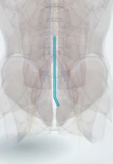
Filum terminale !
The filum terminale is a thin strand running from the end of the spinal cord down to the coccyx; it anchors the lower end of the cord.
Official name: Filum terminale internum
Opens in the Durgical app, no sign-up needed.
What it is
A thin strand that continues from the end of the spinal cord down to the coccyx.
What it does
Anchors the lower end of the cord. If it is too tight from birth (tethered cord), it can stretch the cord.
Key facts
- Where
- From the conus (L1–L2): filum internum inside the dural sac to S2 (about 15 cm), filum externum below S2 (about 5 cm); about 20 cm in all
- Attaches
- Conus medullaris above; dorsum of the first coccygeal segment below (as the coccygeal ligament)
- Function
- Pial strand that anchors the lower end of the cord and dural sac
Relations
- Above
- The tip of the conus medullaris
- Lateral
- The roots of the cauda equina, floating around it in the lumbar cistern
- Below
- After leaving the end of the dural sac at about S2, it runs through the sacral canal to the back of the first coccygeal segment
How to find it
It cannot be felt. It runs from the tip of the conus (usually at L1–L2) down the middle of the cauda equina, leaves the dural sac at about S2 and ends on the back of the first coccygeal segment; for detethering it is reached through a short laminectomy low in the lumbar spine.
On imaging
Axial and sagittal T1 MRI show a fatty filum as a bright strand; its thickness is measured on axial images at about L5–S1. In young infants, ultrasound shows the filum and whether the conus and roots move freely.
Clinical notes
- Tethered cord symptoms often appear in growth spurts: back and leg pain worse on bending, leg weakness or numbness, a progressive cavus foot or scoliosis, and bladder trouble
- Skin markers over the lower back (hairy patch, a dimple high above the gluteal cleft, haemangioma, fatty lump) suggest a hidden spinal dysraphism with a tethered cord
- Myxopapillary ependymoma is the typical tumour of the filum and conus, in young adults, causing back pain and sometimes cauda equina symptoms
Anatomical variants
- Some fat in the filum is found on MRI in a small proportion of healthy adults; it matters only with a low conus or symptoms
Development
The filum is the remnant of the tail end of the neural tube, left behind as the column outgrows the cord; faulty regression leaves a thick or fatty filum.
Related conditions
Test yourself
Think of the answer, then tap the question to see it.
What are the attachments of the filum terminale?
From the conus medullaris above to the back of the first coccygeal segment below, as the coccygeal ligament after leaving the dural sac at about S2.
Which filum terminale finding is the classic cause of tethered cord syndrome?
A thickened filum (over about 2 mm) or a fatty filum that holds the conus medullaris low.
Which tumour is typical of the filum terminale and conus?
Myxopapillary ependymoma, in young adults, causing back pain and sometimes cauda equina symptoms.
Educational information, not medical advice. For any symptom, see a health professional.
Its names
- Official Latin (TA2)
- filum terminale internum; filum terminale externum TA2 5414
- Official Spanish (DeCS)
- Filum terminal DeCS D002420
- English (TA2)
- Filum terminale internum
- Also called
- pial part of filum terminale · pial filament · dural part of filum terminale · coccygeal ligament · terminal filum · filamentum terminale · filum terminale pialis
Related
Sources: Terminologia Anatomica (TA2); DeCS; Neuroanatomy, Conus Medullaris.
Spotted a mistake?We are always improving for the community. Tell us what is wrong and our team will review it.Report a mistake
Representative model, for educational purposes only.