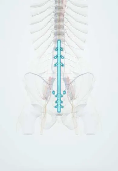
Cauda equina !
The cauda equina is the bundle of nerve roots, like a horse's tail, that fills the lower spinal canal; it serves the legs, bladder and bowel.
Opens in the Durgical app, no sign-up needed.
What it is
The bundle of nerve roots that fills the lower spinal canal below the end of the cord, like a horse's tail.
What it does
Carries the nerves for the legs, bladder, bowel and sexual function down to their exits from the spine.
Key facts
- Roots
- Dorsal and ventral roots of L2–Co1 below the conus, floating in the lumbar cistern (CSF) inside the dural sac
- Moves
- Lower limb muscles; pelvic floor; external anal and urethral sphincters (S2–S4)
- Feeling from
- Lower limbs; saddle (perineal) area; bladder and rectum; cauda equina syndrome: saddle anaesthesia, retention
Relations
- Course
- Each root runs down inside the dural sac, leaves it in a dural sleeve just above its own foramen and exits below the pedicle of its own vertebra; the sac ends at about S2
- Above
- The conus medullaris, from which the lower roots emerge at about L1–L2
- In front
- The posterior longitudinal ligament and the lumbar discs, the source of central herniations
- Behind
- Ligamenta flava and laminae; a thickened ligament and arthritic facet joints narrow the canal from behind
- Contents
- Paired dorsal and ventral roots of L2 to Co1 with the filum terminale in the middle, all in the CSF of the lumbar cistern
Examination
- Perianal pinprick and light touch (S3–S5) with a rectal examination for anal tone and voluntary squeeze: loss supports cauda equina syndrome
- Bladder scan after voiding: a large residual volume shows retention
- Anal wink and bulbocavernosus reflexes (S2–S4): absent with sacral root damage
- Both legs examined for myotomes, ankle reflexes and straight-leg raise; findings on both sides are a warning sign
On imaging
Urgent lumbar MRI is the study of choice, with CT myelography when MRI is not possible. On axial images the roots are dots in the dural sac; clumped roots suggest arachnoiditis, and long curling roots above a tight stenosis show severe narrowing.
Common problems
- Cauda equina syndrome: a large central disc herniation or other mass squeezes the whole bundle. Warning signs: numbness in the 'saddle' area, new bladder or bowel trouble, weakness in both legs. It is a surgical emergency.
- Spinal stenosis can crowd the bundle and cause leg symptoms with walking.
How it is diagnosed
Urgent MRI when warning signs appear.
Treatment
- Emergency decompression surgery (discectomy or laminectomy) to protect nerve function.
Clinical notes
- Lumbar spinal stenosis gives neurogenic claudication: buttock and leg pain or heaviness on walking or standing, eased by sitting or leaning forward; unlike vascular claudication, foot pulses are normal
- Spinal anaesthesia is given into the lumbar cistern among the roots; a needle touching a root causes a brief shooting pain into the leg
- Arachnoiditis after surgery, infection or intrathecal injections can stick the roots together and cause chronic burning leg pain
Development
Until about the third fetal month the cord fills the canal and the roots leave at right angles. The column then grows faster than the cord, whose lower end rises, so the lumbar and sacral roots lengthen downward inside the sac to reach their own foramina, forming the cauda equina.
Related conditions
Test yourself
Think of the answer, then tap the question to see it.
Which roots form the cauda equina?
The dorsal and ventral roots of L2 to Co1 below the conus, floating in the CSF of the lumbar cistern inside the dural sac.
What is the commonest cause of cauda equina syndrome?
A large central disc herniation at L4–L5 or L5–S1; others are tumours, epidural abscess or haematoma, fractures and severe stenosis.
What are the warning signs of cauda equina syndrome?
Saddle numbness, new bladder or bowel trouble (retention) and weakness in both legs; it is a surgical emergency needing urgent MRI.
Why does the timing of decompression matter in cauda equina syndrome?
Outcome is better when decompression comes before urinary retention sets in (incomplete, CES-I) than after (CES-R).
How does neurogenic claudication from lumbar stenosis differ from vascular claudication?
Neurogenic pain eases with sitting or leaning forward, and foot pulses are normal.
Educational information, not medical advice. For any symptom, see a health professional.
Its names
- Official Latin (TA2)
- cauda equina TA2 6163
- Official Spanish (DeCS)
- Cauda equina DeCS D002420
- English (TA2)
- Cauda equina
Related
- Conus medullaris
- Dura (dural sac)
- Filum terminale
- L4 nerve root
- L5 nerve root
- S1 nerve root
- Lumbar arteries
Sources: Terminologia Anatomica (TA2); DeCS; Anatomy, Back, Cauda Equina; 13.2 The Central Nervous System (OpenStax (Rice University), 2022) · adapted, CC BY 4.0; Anatomy of the Human Body (20th ed.): Development of the Nervous System (Lea & Febiger (Gray's Anatomy, 1918), 1918) · adapted, Public domain.
Spotted a mistake?We are always improving for the community. Tell us what is wrong and our team will review it.Report a mistake
Representative model, for educational purposes only.