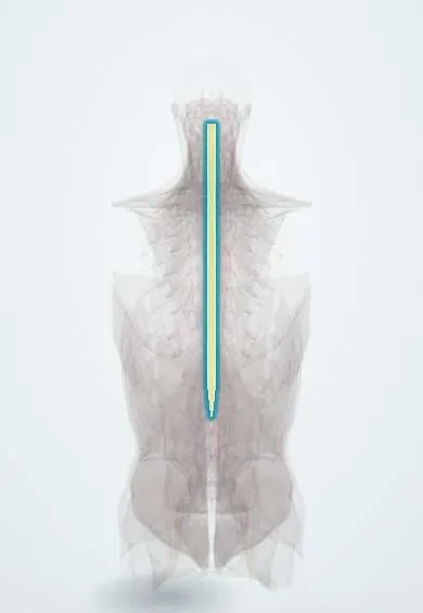
Spina bifida (myelomeningocele) !
Spina bifida (myelomeningocele) is a birth defect where the spine does not close and the spinal cord pushes out in a sac on the back.
Updated 2026-10-29
Structures involved
Tap one to see what it is and open it in 3D.
- Spinal cord
- Dura (dural sac)
- L5 vertebra
- Sacrum
- Cauda equina
- Conus medullaris
- Filum terminale
- L4 vertebra
- Base of the skull
In biochemistry
In embryology
- Germ-layer derivatives: ectoderm, mesoderm, endoderm
- Neurulation and closure of the neural tube
- Vertebrae: resegmentation of the sclerotomes
- Clubfoot (talipes equinovarus)
- Hemivertebra, congenital scoliosis and Klippel–Feil
- Spinal cord: alar and basal plates and the ascent of the cord
- Neural tube defects: anencephaly, spina bifida and encephalocele
- Chiari malformations
- Prenatal diagnosis: ultrasound, serum screening, cell-free DNA, CVS, amniocentesis
- Folic acid and neural tube defect prevention
- Valproate, carbamazepine and phenytoin
Why it happens
What it is
Neural tube defect in which the vertebral arches fail to close and the spinal cord and its coverings protrude through an open sac on the back. It is the most severe form of open spina bifida.
Who gets it
One of the most common congenital defects of the nervous system. Rates have fallen in countries that fortify flour with folic acid, including several in Latin America.
Causes
- Failure of the caudal neural tube to close in the first month of pregnancy
- Multifactorial: genetic and environmental factors combined
Risk factors
- Low maternal folate
- Previous pregnancy with a neural tube defect or a family history
- Maternal diabetes and obesity
- Some antiseizure drugs in pregnancy, such as valproate
- Maternal fever or hyperthermia in early pregnancy
Pathophysiology
The exposed neural tissue is malformed and is further damaged in the womb by amniotic fluid and trauma. Loss of function below the level causes weakness and sensory loss of the legs and neurogenic bladder and bowel. Leakage of cerebrospinal fluid pulls the hindbrain downward (Chiari II malformation), which blocks fluid flow and causes hydrocephalus.
Classification
- Spina bifida occulta: closed bony defect, often with no symptoms
- Meningocele: sac with meninges and fluid but no neural tissue
- Myelomeningocele: sac containing spinal cord or nerve roots
- By level: thoracic, lumbar or sacral; higher lesions cause more deficit
Symptoms and signs
- Open sac on the back, most often lumbosacral, seen at birth or on prenatal ultrasound
- Leg weakness or paralysis and loss of sensation below the level
- Foot and hip deformities (clubfoot, hip dislocation)
- Neurogenic bladder and bowel
- Signs of hydrocephalus: large head, bulging fontanelle
- Brainstem signs from Chiari II: feeding problems, stridor, apnoea
Diagnosis
- Clinical
- Inspection of the back at birth and full neurological exam to define the functional level; head circumference and fontanelle checks.
- Tests
- Maternal serum alpha-fetoprotein screening in pregnancy (raised in open defects). Urodynamic studies and renal function after birth.
- Imaging
- Prenatal ultrasound shows the defect and skull signs (lemon sign) and cerebellar signs (banana sign); fetal MRI adds detail. After birth: head ultrasound or MRI for hydrocephalus and Chiari II, spine MRI, and kidney ultrasound.
Differential diagnosis
- Meningocele or lipomyelomeningocele
- Sacrococcygeal teratoma
- Spina bifida occulta
- Other congenital spinal masses
Goal of treatment
Close the defect to prevent infection and further damage, control hydrocephalus, and support lifelong function of the kidneys, bladder, bowel, limbs and learning.
Treatment
- Surgical
- Surgical closure of the defect within the first days after birth, with the sac kept moist and covered and infection prevented until then; fetal repair in selected pregnancies at specialised centres reduces the need for a shunt.
- Surgical
- Treatment of hydrocephalus with a ventriculoperitoneal shunt or endoscopic third ventriculostomy.
- Follow-up
- Lifelong follow-up by a team: urology (clean intermittent catheterisation, bladder medicines), bowel programme, kidney checks, and surveillance for tethered cord and scoliosis.
- Rehabilitation
- Rehabilitation and orthopaedics: physiotherapy, braces and mobility aids, care of the feet and hips, and school and psychosocial support.
Drugs for this condition
Complications
- Shunt failure or infection
- Urinary tract infections and kidney damage
- Tethered spinal cord
- Scoliosis and hip dislocation
- Latex allergy
- Pressure sores and fractures in areas without sensation
- Learning difficulties
Prognosis
Most children survive into adulthood with good care. Function depends on the level of the lesion and on hydrocephalus; kidney protection and shunt care are key to long-term health.
Prevention
- Folic acid supplementation for all women who could become pregnant, starting before conception (higher supplementation after an affected pregnancy, as guidelines set)
- Folic acid fortification of staple foods such as flour, a proven public-health measure
- Control of maternal diabetes and review of medications before pregnancy
Test yourself
Think of the answer, then tap the question to see it.
Which public-health measure has reduced the frequency of neural tube defects?
Folic acid supplementation before conception and fortification of staple foods such as flour with folic acid.
Which brain malformation is almost always associated with myelomeningocele?
Chiari II malformation, with downward displacement of the hindbrain.
Why do many children with myelomeningocele develop hydrocephalus?
The Chiari II malformation blocks the flow of cerebrospinal fluid out of the brain's ventricles.
What do the lemon and banana signs on prenatal ultrasound suggest?
Open spina bifida: a lemon-shaped skull and a banana-shaped cerebellum from the Chiari II malformation.
Why are children with spina bifida managed in a latex-free environment?
They have a high risk of latex allergy from repeated early exposure during surgery and catheterisation.
Related conditions
- Scoliosis
- Developmental dysplasia of the hip
- Urinary tract infection (acute cystitis)
- Clubfoot (talipes equinovarus)
- Hydrocephalus
Educational information, not medical advice. For any symptom, see a health professional.
Sources: Myelomeningocele (StatPearls (NCBI Bookshelf), 2024).
Spotted a mistake?We are always improving for the community. Tell us what is wrong and our team will review it.Report a mistake
Representative model, for educational purposes only.