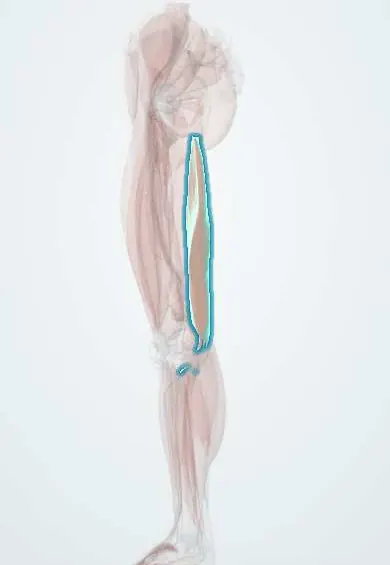
Semimembranosus !
The semimembranosus is the deepest and broadest inner hamstring muscle at the back of the thigh; it bends the knee and extends the hip.
Official name: Semimembranosus muscle
Opens in the Durgical app, no sign-up needed.
What it is
The deepest inner hamstring muscle, at the back of the thigh.
What it does
Bends the knee and extends the hip; helps control rotation of the knee.
Key facts
- Origin
- Ischial tuberosity (superolateral impression)
- Insertion
- Posterior medial tibial condyle (direct arm); medial tibial condyle under the MCL (reflected arm); recurrent expansion forms the oblique popliteal ligament
- Action
- Extends hip; flexes knee; medially rotates flexed knee
- Nerve
- Sciatic nerve, tibial division (L5–S2)
- Blood supply
- Perforating arteries; medial circumflex femoral artery; popliteal artery branches
Relations
- Superficial
- Semitendinosus, lying in a groove on its back surface
- Deep
- Adductor magnus above; at the knee, the medial head of the gastrocnemius and the posteromedial joint capsule, with the semimembranosus–gastrocnemius bursa between them
- Lateral
- Sciatic nerve in the thigh; lower down, the tibial nerve and the popliteal artery and vein in the popliteal fossa
- Medial
- Gracilis, and the sartorius near the knee
- Below
- Arms to the posteromedial capsule, the popliteus fascia and, in many descriptions, the posterior horn of the medial meniscus, which it draws back as the knee bends
How to find it
Prone with the knee bent against resistance, it is felt as a soft, fleshy mass on both sides of and deep to the semitendinosus tendon, best just above the inner back of the knee.
Examination
- Resisted knee flexion with the leg turned inward: pain at the posteromedial knee or in the medial hamstring belly suggests a semimembranosus strain or insertional tendinopathy
On imaging
On axial MRI or ultrasound of the knee, a popliteal (Baker) cyst is a fluid collection whose neck passes between the semimembranosus tendon and the medial head of the gastrocnemius; this neck tells it apart from other masses of the popliteal fossa.
Common problems
- Hamstring strain, common in sprinting sports.
- Rarely, the tendon pulls off the bone at the hip.
How it is diagnosed
Physical exam; MRI for severe strains.
Treatment
- Rest, then progressive stretching and strengthening; surgery only for complete tendon pull-offs.
Clinical notes
- Insertional tendinopathy causes posteromedial knee pain that can mimic a tear of the medial meniscus
- Injuries of the posteromedial corner that come with anterior cruciate or medial collateral ligament tears often involve its capsular arms and add rotational instability
Development
From the ventral (flexor) muscle mass, and therefore supplied by the tibial division of the sciatic nerve.
Related conditions
Test yourself
Think of the answer, then tap the question to see it.
Between which two structures does a popliteal (Baker) cyst arise?
In the bursa between the semimembranosus tendon and the medial head of the gastrocnemius; the cyst's neck passes between them on MRI or ultrasound.
Which knee ligament is formed by an expansion of the semimembranosus tendon?
The oblique popliteal ligament, which strengthens the back of the knee capsule.
What role does the distal semimembranosus tendon play at the knee?
It splits into several arms around the posteromedial knee and is a key active stabilizer of the posteromedial corner.
Which knee problem can semimembranosus insertional tendinopathy mimic?
A tear of the medial meniscus, because both cause posteromedial knee pain.
Educational information, not medical advice. For any symptom, see a health professional.
Its names
- Official Latin (TA2)
- musculus semimembranosus TA2 2642
- Official Spanish (DeCS)
- Músculo semimembranoso DeCS D000070633
- English (TA2)
- Semimembranosus muscle
- Also called
- musculus semimembranaceus
Related
Sources: Terminologia Anatomica (TA2); DeCS; Anatomy, Bony Pelvis and Lower Limb, Hamstring Muscle; Anatomy, Bony Pelvis and Lower Limb: Posterior Thigh Muscles.
Spotted a mistake?We are always improving for the community. Tell us what is wrong and our team will review it.Report a mistake
Representative model, for educational purposes only.