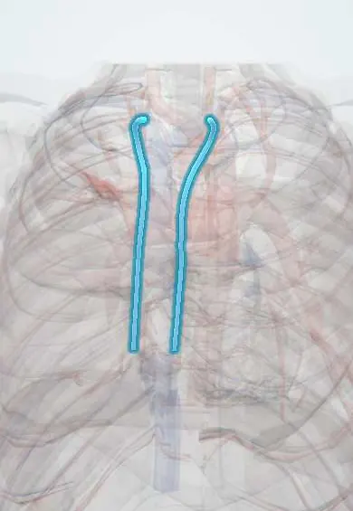
Internal thoracic veins !
The internal thoracic veins run beside the artery of the same name inside the front chest wall and drain it into the brachiocephalic veins.
Opens in the Durgical app, no sign-up needed.
What it is
Veins running with the internal thoracic artery.
What it does
Drain the front chest wall into the brachiocephalic veins.
Key facts
- Drains
- Anterior chest wall and intercostal spaces, breast (perforating veins); superior epigastric and musculophrenic veins
- Into
- Brachiocephalic vein of same side
Relations
- Course
- From the musculophrenic and superior epigastric veins at the 6th space they ascend behind the costal cartilages beside the artery, collect anterior intercostal and perforating veins, and end in the brachiocephalic vein of the same side
- In front
- Costal cartilages and the front ends of the internal intercostal muscles
- Behind
- Endothoracic fascia and pleura above; the transversus thoracis muscle below
- Lateral
- The internal thoracic artery, where the single upper vein lies on its medial side
- Medial
- Sternal edge and the parasternal lymph nodes
How to find it
Not palpable; they follow the internal thoracic artery about a finger-breadth from the sternal edge and are seen beside it on contrast CT and on ultrasound.
On imaging
Seen on contrast CT beside the artery, behind the costal cartilages next to the sternum; along this line internal mammary nodes are looked for in breast cancer staging. In superior vena cava obstruction CT shows them and the chest-wall veins enlarged as collaterals. Before free-flap breast reconstruction, CT angiography or Doppler checks their size.
Common problems
- Rarely diseased.
Treatment
- In bypass surgery the left internal mammary artery is freed and joined to the LAD; it often stays open for decades.
Clinical notes
- Taken with the artery in a pedicled coronary graft, or left in place in a skeletonized harvest
- Bleeding from them or from side branches is one source of bleeding after a sternotomy
- They dilate, with the chest-wall veins, in superior vena cava obstruction
Test yourself
Think of the answer, then tap the question to see it.
Into which vein do the internal thoracic veins drain?
The brachiocephalic vein of the same side, after the two veins beside the artery merge into one vein medial to it.
How do the internal thoracic veins link the superior and inferior vena cava systems?
Through the superior and inferior epigastric veins, a collateral path that enlarges when either cava is blocked.
In which reconstructive operation are the internal thoracic veins the recipient veins of choice?
Free-flap breast reconstruction, together with the internal thoracic artery.
Educational information, not medical advice. For any symptom, see a health professional.
Its names
- Official Latin (TA2)
- venae thoracicae internae TA2 4786
- Official Spanish (DeCS)
- Venas torácicas internas
- English (TA2)
- Internal thoracic veins
- Also called
- vena mammaria interna
Sources: Terminologia Anatomica (TA2); Anatomy, Thorax, Internal Mammary (Internal Thoracic) Arteries.
Spotted a mistake?We are always improving for the community. Tell us what is wrong and our team will review it.Report a mistake
Representative model, for educational purposes only.