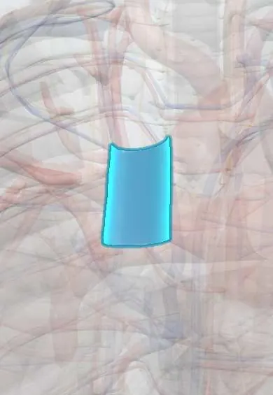
Superior vena cava !
The superior vena cava is the large vein that returns oxygen-poor blood from the head, neck, arms and upper chest to the right atrium.
Opens in the Durgical app, no sign-up needed.
What it is
The large vein bringing blood from the head, neck and arms into the right atrium.
What it does
Returns oxygen-poor blood from the upper body. Pacemaker leads and central lines pass through it.
Key facts
- Drains
- Head, neck, upper limbs and chest wall, via both brachiocephalic veins and azygos vein; formed behind right 1st costal cartilage
- Into
- Right atrium at level of right 3rd costal cartilage; valveless
Relations
- Course
- Formed behind the right 1st costal cartilage, it descends vertically along the right sternal border, receives the azygos vein at the 2nd costal cartilage level and enters the right atrium at the 3rd
- In front
- Anterior margins of the right lung and pleura, the thymic remnant, and the right 1st and 2nd costal cartilages and intercostal space
- Behind
- Trachea and right vagus nerve above; the right lung root (right pulmonary artery and right main bronchus) below
- Medial
- Ascending aorta and brachiocephalic trunk
- Lateral
- Right phrenic nerve with the pericardiacophrenic vessels, then the mediastinal pleura and right lung
How to find it
Not palpable. Its line on the chest wall runs along the right edge of the sternum, from behind the right 1st costal cartilage, where the two brachiocephalic veins join, down to the right 3rd costal cartilage, where it opens into the right atrium.
On imaging
On a frontal chest X-ray it forms the upper right mediastinal border. The carina is used as a landmark for central catheter tips, which are aimed at the lower cava or its junction with the right atrium. Contrast CT shows obstruction, thrombus and collateral veins.
Common problems
- Superior vena cava syndrome: blockage by a tumour or clot, causing swelling of the face and arms.
Clinical notes
- Superior vena cava syndrome is caused mostly by lung cancer and then lymphoma; clots around catheters and pacemaker leads are an increasing cause
- Signs: swollen face and arms, distended neck and chest-wall veins, worse lying flat or bending forward; raising both arms can bring on facial plethora (Pemberton sign)
- Route for central venous catheters, pacemaker and defibrillator leads and right heart catheterization from the neck or arm
- Cannulated for cardiopulmonary bypass; stitches or clamps near the cavoatrial junction can damage the sinoatrial node
Anatomical variants
- Persistent left superior vena cava in about 0.3% of people (more often with congenital heart disease), usually draining into an enlarged coronary sinus; rarely it opens into the left atrium, a right-to-left shunt that can carry paradoxical emboli
- Double superior vena cava (right and persistent left), with or without a bridging left brachiocephalic vein
Development
Forms from the right anterior cardinal vein and the right common cardinal vein; the left-sided equivalents normally regress, leaving the coronary sinus and the oblique vein of the left atrium.
Related conditions
Test yourself
Think of the answer, then tap the question to see it.
Where is the superior vena cava formed, and where does it enter the right atrium?
It forms behind the right 1st costal cartilage and enters the right atrium at the level of the right 3rd costal cartilage.
Which vein joins the superior vena cava from behind, and at what level?
The azygos vein, arching over the right lung root at the sternal angle level (T4–T5).
What are the main causes of superior vena cava syndrome?
Mostly lung cancer, then lymphoma; clots around catheters and pacemaker leads are an increasing cause.
What is the Pemberton sign in superior vena cava obstruction?
Facial plethora brought on by raising both arms.
Which conduction structure lies at the junction of the superior vena cava with the right atrium?
The sinoatrial node, at the top of the crista terminalis; stitches or clamps near the cavoatrial junction can damage it.
Educational information, not medical advice. For any symptom, see a health professional.
Its names
- Official Latin (TA2)
- vena cava superior TA2 4745
- Official Spanish (DeCS)
- Vena cava superior DeCS D014683
- English (TA2)
- Superior vena cava
- Also called
- vena cava cranialis
Related
Sources: Terminologia Anatomica (TA2); DeCS; Anatomy, Thorax, Superior Vena Cava; Anatomy of the Human Body (20th ed.): Surface Markings of the Thorax (Lea & Febiger (Gray's Anatomy, 1918), 1918) · adapted, Public domain; Tratado de anatomía humana (9.ª ed.), tomo II, Libro IV Angiología, sección 4: Venas (sistemas de las venas cavas superior e inferior, vena porta, venas del raquis).
Spotted a mistake?We are always improving for the community. Tell us what is wrong and our team will review it.Report a mistake
Representative model, for educational purposes only.