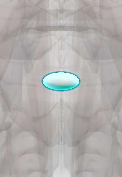
L5–S1 nucleus !
The nucleus pulposus is the soft, gel-like centre of the L5-S1 disc in the lower back; it is the part that bulges out in a disc herniation.
Official name: L5-S1
Opens in the Durgical app, no sign-up needed.
What it is
The soft, gel-like centre (nucleus pulposus) of the L5–S1 disc.
What it does
Works like a water cushion: it turns pressure from above into outward push on the fibre ring, spreading the load. It is under the most pressure when you bend forward and lift.
Key facts
- Where
- Centre of the L5–S1 disc, slightly toward the back, inside the annulus fibrosus
- Function
- Notochord-derived gel (about 70–90% water, proteoglycans); turns axial load into annulus tension; herniates posterolaterally toward the S1 root
Relations
- In front
- Below the aortic bifurcation: between the common iliac vessels lie the median sacral vessels and the superior hypogastric plexus, whose injury in anterior approaches can cause retrograde ejaculation
- Behind
- Posterior longitudinal ligament and the dural sac; the S1 root runs in the lateral recess
- Lateral
- The L5 root in its foramen, which is often narrowed at this level
How to find it
From behind, the L5–S1 interlaminar window lies between the L5 spinous process and the back of the sacrum and is the widest in the lumbar spine. From the front, the disc sits just below the sacral promontory, between the common iliac vessels.
Examination
- S1 root: weak toe walking and calf raises, numb outer foot and sole, reduced or absent ankle jerk
On imaging
Sagittal T2 MRI follows a migrated fragment up or down; a dark, thin L5–S1 disc with a «vacuum» (gas) on CT or X-ray marks advanced degeneration.
Common problems
- Squeezes out through a tear in the ring (herniation), usually backward and to one side, toward a nerve root.
- Loses water with age, so the disc gets thinner and stiffer.
How it is diagnosed
On MRI a healthy nucleus is bright on T2 images; a worn one turns dark ('black disc').
Treatment
- In a microdiscectomy only the loose piece that has pushed out is removed; most of the disc stays.
Clinical notes
- L4–L5 and L5–S1 together account for most lumbar herniations; a posterolateral L5–S1 fragment usually takes the S1 root, a far-lateral one the L5 root
- In isthmic spondylolisthesis of L5 on S1 the disc wears and the slip pinches the L5 root in its foramen, rather than the S1 root a herniation usually hits
- Because the interlaminar window is widest here, an L5–S1 fragment can often be removed with little or no bone taken from the lamina
Anatomical variants
- Lumbosacral transitional vertebrae (a sacralised L5 or a lumbarised S1) are common and change the numbering, so the disc called L5–S1 on one report may be named differently on another; levels are checked against a whole-spine image
Development
The nucleus pulposus comes from the notochord; notochordal cells give way to cartilage-like cells during childhood. Notochord remnants in the sacrum are the usual source of sacral chordoma.
Related conditions
Test yourself
Think of the answer, then tap the question to see it.
Toward which root does a posterolateral herniation of the L5–S1 nucleus usually go?
The S1 root, from a fragment escaping backward and to one side.
Why can a central L5–S1 nucleus herniation cause cauda equina syndrome with less leg pain than expected?
The lower sacral roots for bladder, bowel and saddle lie centrally in the dural sac, so a central fragment hits them first.
What decides which root a sequestered L5–S1 nucleus fragment compresses?
Where it ends up: it can migrate up behind L5 or down behind S1.
Which tumour arises from notochord remnants in the sacrum, near the L5–S1 nucleus?
Sacral chordoma; the nucleus pulposus itself comes from the notochord.
Educational information, not medical advice. For any symptom, see a health professional.
Its names
- Official Latin (TA2)
- nucleus pulposus L5-S1 TA2 1686
- Official Spanish (DeCS)
- Núcleo pulposo L5-S1 DeCS D000070614
- English (TA2)
- L5-S1
Related
Sources: Terminologia Anatomica (TA2); DeCS; Anatomy, Back, Nucleus Pulposus; Anatomy, Back, Intervertebral Discs; 7.3 The Vertebral Column (OpenStax (Rice University), 2022) · adapted, CC BY 4.0.
Spotted a mistake?We are always improving for the community. Tell us what is wrong and our team will review it.Report a mistake
Representative model, for educational purposes only.