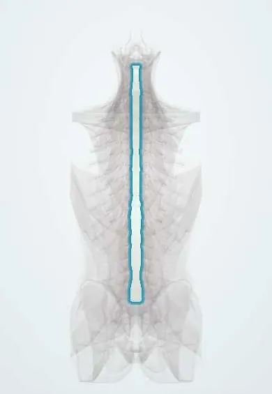
Posterior longitudinal ligament !
The posterior longitudinal ligament runs down the back of the vertebral bodies and discs, inside the spinal canal, in front of the cord.
Opens in the Durgical app, no sign-up needed.
What it is
A narrower band running down the back of the vertebral bodies and discs, inside the spinal canal, in front of the spinal cord.
What it does
Limits forward bending and covers the back of the discs, which is one reason most herniations go off to the side rather than straight back.
Key facts
- Attaches
- Back of vertebral bodies and discs inside the canal, from C2 (continues up as tectorial membrane) to the sacrum (some texts: down to the base of the coccyx)
- Function
- Limits flexion; resists posterior disc herniation; narrow centrally (narrowest in lumbar), so herniations go posterolateral
- Tight in
- Flexion (bending forward)
Relations
- In front
- Back of the vertebral bodies and discs; beneath it, the basivertebral veins leave the bodies and join the anterior internal vertebral venous plexus
- Behind
- Anterior epidural space, then the dural sac with the spinal cord or the cauda equina; fine meningovertebral ligaments tether the dura to it
- Lateral
- Its edges leave the posterolateral corner of each disc uncovered, next to the lateral recess and the intervertebral foramen
How to find it
Not palpable. In anterior cervical discectomy it is the last layer reached once the disc is cleared, a white band lying on the dura. From behind it is seen only by gently moving the dural sac or root aside, as in lumbar discectomy.
On imaging
MRI shows it as a thin dark line behind the bodies and discs, and shows cord compression and cord signal change. CT shows OPLL as a bony plate behind the bodies, typed as continuous, segmental, mixed or circumscribed. Sagittal CT is the best view for its length and thickness.
Common problems
- OPLL: the ligament slowly turns to bone and narrows the canal in the neck (more common in East Asian populations).
- Disc herniations push against it, or through it.
How it is diagnosed
CT shows ossification; MRI shows cord compression.
Treatment
- Surgery to take pressure off the cord if it is pressed. In an ACDF the surgeon often removes it at that level to free the cord and nerve roots.
Clinical notes
- In burst fractures, an intact ligament allows distraction to pull retropulsed fragments back out of the canal (ligamentotaxis)
- In anterior cervical discectomy it is opened or removed to find fragments that have passed behind it
- In OPLL with an ossified dura, removing the bone from the front risks a CSF leak; posterior approaches are often chosen for long, multilevel disease
Related conditions
Test yourself
Think of the answer, then tap the question to see it.
Where does the posterior longitudinal ligament run?
Down the back of the vertebral bodies and discs inside the canal, from C2 (continuing up as the tectorial membrane) to the sacrum.
Which movement does the posterior longitudinal ligament limit?
Flexion (bending forward); it is taut in flexion.
Why do most lumbar disc herniations go posterolateral rather than straight back?
The posterior longitudinal ligament is narrow centrally and thinnest at L5–S1, and its edges leave the posterolateral corner of each disc uncovered.
What is OPLL, and what does a «double-layer sign» on CT suggest?
Ossification of the posterior longitudinal ligament, mostly mid-cervical and in East Asian populations, causing myelopathy; the double-layer sign suggests the dura is ossified too.
In a burst fracture, how does an intact posterior longitudinal ligament help reduction?
Ligamentotaxis: distraction tightens the intact ligament, which pulls retropulsed fragments back out of the canal.
Educational information, not medical advice. For any symptom, see a health professional.
Its names
- Official Latin (TA2)
- ligamentum longitudinale posterius TA2 1680
- Official Spanish (DeCS)
- Ligamento longitudinal posterior DeCS D017846
- English (TA2)
- Posterior longitudinal ligament
- Also called
- PLL · ligamentum longitudinale commune dorsale
Related
- Anterior longitudinal ligament
- Dura (dural sac)
- Axis (C2)
- C3 vertebra
- C4 vertebra
- C5 vertebra
- C6 vertebra
- L1 vertebra
Sources: Terminologia Anatomica (TA2); DeCS; Anatomy, Back, Posterior Longitudinal Ligament.
Spotted a mistake?We are always improving for the community. Tell us what is wrong and our team will review it.Report a mistake
Representative model, for educational purposes only.