Digestive system · Head and neck
Lower left third molar !
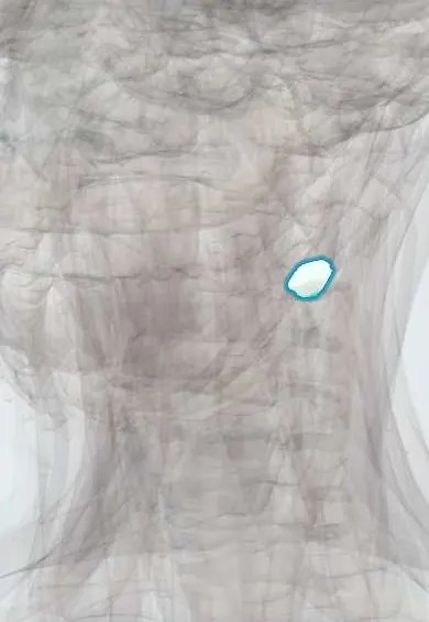
The lower left third molar (tooth 38) is the wisdom tooth, last at the back; it often lacks room to come in and stays stuck (impacted).
Opens in the Durgical app, no sign-up needed.
What it is
The wisdom tooth: the last molar, usually coming in between the ages of 17 and 25.
What it does
Grinds food when it erupts fully; often it does not.
Key facts
- Joints with
- Alveolar socket of the mandible (gomphosis held by the periodontal ligament); bites against the opposing teeth
- Landmarks
- FDI 38, lower left third molar (wisdom tooth): roots often fused or variable; often impacted or missing; erupts at about 17–25 years; nerve: inferior alveolar nerve (V3); close to the inferior alveolar canal and the lingual nerve
Relations
- In front
- The lower left second molar
- Behind
- Front edge of the ramus of the mandible and the retromolar area
- Above
- The upper left second molar and the upper left third molar, when erupted
- Below
- Inferior alveolar canal with the inferior alveolar nerve, artery and vein, often touching the root tips
- Medial
- Lingual nerve inside the thin lingual plate; lower down, the submandibular space below the mylohyoid line
How to find it
The last tooth on the lower left, eighth from the midline, against the front of the ramus; when partly erupted only a cusp shows through a flap of gum.
On imaging
Panoramic radiograph signs of a close canal: darkening of the root where the canal crosses it, a break in the canal's white lines or a diverted canal; cone-beam CT then shows the exact relation.
Common problems
- Impacted wisdom tooth: stuck under the gum or against the second molar, with pain, swelling or infection (pericoronitis).
Clinical notes
- Pericoronitis starts under the gum flap (operculum) over a partly erupted crown; spread into the chewing-muscle spaces brings trismus
- Pell and Gregory grade the impacted tooth by the room left by the ramus (I–III) and its depth against the second molar (A–C)
- Coronectomy, taking off the crown and leaving the roots, is an option when the roots are bound to the canal
- Lower third molars are the most common site of a dentigerous cyst, which forms around the crown of an unerupted tooth
Anatomical variants
- Crown and roots vary widely: extra or fewer cusps, and roots that are often fused into one cone or curved sharply
Development
The last teeth to form: calcification begins around 9–10 years and the roots close in the early twenties, so their stage helps estimate age in forensic work.
Memory aid
FDI 38 = quadrant 3 (lower left), tooth 8. In the US Universal system it is #17, where the lower count begins.
Related conditions
Test yourself
Think of the answer, then tap the question to see it.
What does Winter's classification describe for the lower left third molar, and which form is most common?
Its tilt when impacted: mesioangular (the most common), vertical, horizontal or distoangular.
Which nerve on the lingual side is at risk in removal of the lower left third molar, and what is the deficit?
The lingual nerve, against the thin lingual plate: numbness of that side of the tongue and possibly altered taste in its front two thirds.
Which panoramic radiograph signs suggest the roots of the lower left third molar are close to the inferior alveolar canal?
Darkening of the root where the canal crosses it, a break in the canal's white lines or a diverted canal; cone-beam CT then shows the exact relation.
What is a dry socket after removal of the lower left third molar?
Alveolar osteitis: the clot is lost and severe pain starts a few days after extraction.
Educational information, not medical advice. For any symptom, see a health professional.
Related
Sources: Anatomy, Head and Neck, Teeth; Anatomy, Permanent Dentition; Anatomy, Head and Neck, Tooth Eruption; Anatomy, Head and Neck: Alveolar Nerve; 23.3 The Mouth, Pharynx, and Esophagus (OpenStax (Rice University), 2022) · adapted, CC BY 4.0.
Spotted a mistake?We are always improving for the community. Tell us what is wrong and our team will review it.Report a mistake
Representative model, for educational purposes only.