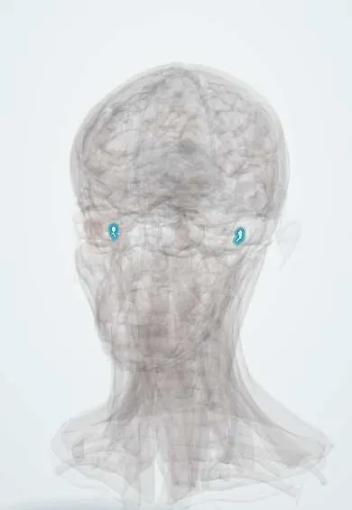
Malleus !
The malleus (hammer) is the first of the three tiny middle ear bones, attached to the eardrum; it passes the eardrum's vibrations on toward the inner ear.
Opens in the Durgical app, no sign-up needed.
What it is
The first ossicle, attached to the eardrum.
What it does
Passes and amplifies the eardrum's vibrations to the inner ear.
Key facts
- Joints with
- Tympanic membrane (handle), incus
- Landmarks
- Head, neck, handle, lateral and anterior processes
Relations
- Lateral
- Tympanic membrane, with the handle embedded in it down to the umbo and the lateral process raising the malleolar prominence
- Behind
- Body of the incus at the incudomalleolar joint, a small synovial saddle joint in the attic
- Above
- Head in the epitympanic recess under the tegmen tympani, the thin roof between the middle ear and the middle cranial fossa
- Medial
- Tendon of the tensor tympani hooking around the cochleariform process; the chorda tympani passing across
- In front
- Anterior process and ligament, toward the petrotympanic fissure through which the chorda tympani leaves the middle ear
How to find it
Through a normal eardrum, the handle is seen as a pale streak running from the lateral process (a small white knob at the top) down and back to the umbo at the centre. The head is hidden in the attic and is reached surgically after removing bone of the outer attic wall.
Examination
- Tympanometry: a low, shallow peak (type As) with a conductive loss suggests a stiff chain, as in fixation of the malleus head or otosclerosis; an unusually tall peak (type Ad) suggests a broken chain
- Rinne and Weber: a conductive pattern (Rinne negative, Weber toward that ear) behind a normal-looking drum points to ossicular fixation or a break in the chain
On imaging
High-resolution temporal bone CT shows it best: on axial images its head and the body of the incus form an «ice cream cone» in the attic. Look for erosion of the head by an attic cholesteatoma, fixation in tympanosclerosis, and fracture or separation after a temporal bone injury. On otoscopy its handle and lateral process are seen through the drum.
Clinical notes
- Attic cholesteatoma erodes the head of the malleus and the body of the incus; these are often removed at surgery and the chain rebuilt
- Ossiculoplasty connects the malleus handle or the eardrum to the stapes with a prosthesis or the patient's own reshaped incus
- Fixation of the malleus head in the attic (from tympanosclerosis or bony bridging) causes conductive loss with a normal-looking drum
- A shortened, more horizontal-looking handle with a prominent lateral process is a sign of a retracted drum from negative middle ear pressure
Development
Malleus and incus come mainly from the first pharyngeal arch (Meckel cartilage); the ossicles are already adult-sized at birth.
Memory aid
From outside in, «MIS»: Malleus, Incus, Stapes; the hammer strikes the anvil, which moves the stirrup.
Related conditions
Test yourself
Think of the answer, then tap the question to see it.
Which ossicle is seen through the eardrum, and which part?
The malleus: its handle, running from the lateral process down and back to the umbo.
Which muscle inserts on the malleus, and what is its nerve supply?
The tensor tympani, supplied by the mandibular nerve (V3); it pulls the drum inward and tenses it.
How does the chorda tympani relate to the malleus?
It crosses medial to the handle of the malleus and lateral to the long limb of the incus.
Educational information, not medical advice. For any symptom, see a health professional.
Its names
- Official Latin (TA2)
- malleus TA2 881
- Official Spanish (DeCS)
- Martillo DeCS D008307
- English (TA2)
- Malleus
- Also called
- hammer
Related
Sources: Terminologia Anatomica (TA2); DeCS; Ear, Hearing, and Facial Nerve Evaluation; Physiology, Ear.
Spotted a mistake?We are always improving for the community. Tell us what is wrong and our team will review it.Report a mistake
Representative model, for educational purposes only.