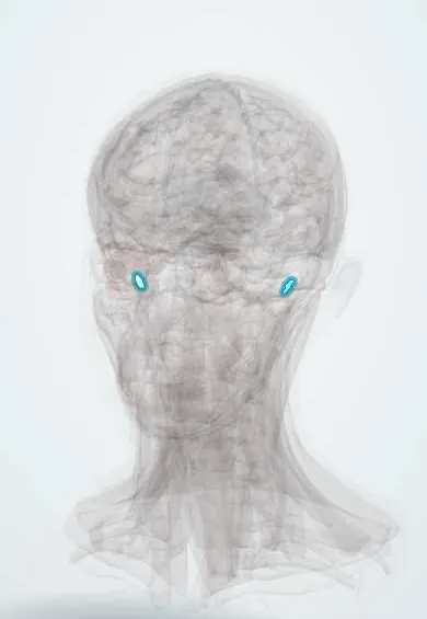
Eardrum !
The eardrum (tympanic membrane) is a thin membrane at the end of the ear canal; it vibrates with sound and passes it to the tiny ear bones.
Official name: Tympanic membrane
Opens in the Durgical app, no sign-up needed.
What it is
A thin cone-shaped membrane at the end of the ear canal.
What it does
Vibrates with sound and passes it to the ossicles; it can burst with infection or pressure.
Key facts
- Where
- End of the ear canal; handle of the malleus attached (umbo); about 1 cm across, tilted about 45° to the horizontal
- Blood supply
- Deep auricular and anterior tympanic arteries
- Nerve
- Outer: V3 and X; inner: IX (tympanic plexus)
- Function
- Converts sound into vibration of the ossicles
Relations
- Lateral
- Deep end of the external acoustic meatus
- Medial
- Middle ear: the handle of the malleus is embedded in it; the chorda tympani crosses its upper inner surface; the promontory and round window lie beyond
- Above
- Behind the pars flaccida, the epitympanic recess (attic) with the head of the malleus and the body of the incus
How to find it
On otoscopy it is pearly grey and translucent and faces down, forward and outward. It is divided into four quadrants by a line along the handle of the malleus and a perpendicular through the umbo; the long process of the incus may show through the posterosuperior quadrant. The cone of light points forward and down (about 5 o'clock in a right ear, 7 o'clock in a left).
Examination
- Pneumatic otoscopy: reduced or absent movement when air pressure is changed shows fluid behind the drum or a perforation
- Tympanometry: type A normal; type B flat with fluid in the middle ear (normal canal volume) or with a perforation or open tube (large volume); type C, negative pressure
- Weber lateralises to the affected ear and Rinne becomes negative with a perforation or effusion: a conductive pattern
On imaging
Examined with the otoscope and pneumatic otoscopy, not with scans; tympanometry tests how it moves (a flat trace in middle ear effusion). On high-resolution temporal bone CT it is a thin line at the end of the canal. CT, or diffusion-weighted MRI, is chosen when a retraction pocket or pars flaccida cholesteatoma is suspected: look for erosion of the scutum and the ossicles.
Common problems
- Middle ear infection (otitis media) makes it red and bulging; it can burst and drain.
- Perforation from infection, a slap, cotton buds or pressure changes.
How it is diagnosed
Looking with an otoscope; a hearing test and tympanometry.
Treatment
- Pain relief; antibiotics when needed.
- Most perforations heal by themselves; surgery (tympanoplasty) if not.
- Ventilation tubes for repeated infections or fluid in children.
Clinical notes
- Acute otitis media: red, bulging drum that hides the landmarks and the cone of light; if it ruptures, pus drains and the pain often eases
- Otitis media with effusion: dull, retracted drum with a fluid level or bubbles; a retracted drum makes the handle of the malleus look short and the lateral process stand out
- Myringotomy and grommets (tympanostomy tubes) use the inferior quadrants, away from the ossicles and chorda tympani (posterosuperior); some texts: posteroinferior to drain, anteroinferior for tubes
- Most small traumatic perforations heal on their own; persistent ones are closed by myringoplasty, often with temporalis fascia or tragal perichondrium
- Tympanosclerosis: chalky white patches after repeated infections or grommets, usually with little hearing effect
Development
It is where the first pharyngeal cleft (outer skin) meets the first pharyngeal pouch (inner mucosa), with mesenchyme between: all three germ layers in one membrane.
Memory aid
The cone of light points toward the face: anteroinferior in both ears, so 5 o'clock on the right and 7 o'clock on the left.
Related conditions
- Acute otitis media
- Hearing loss (conductive and sensorineural, including sudden hearing loss)
- Otitis media with effusion and chronic suppurative otitis media
- Cholesteatoma
Test yourself
Think of the answer, then tap the question to see it.
What are the otoscopic landmarks of the tympanic membrane?
The handle of the malleus running down and back to the umbo, its lateral process at the top, and the cone of light anteroinferiorly.
Which tympanic membrane perforations are «unsafe», and why?
Marginal and attic (pars flaccida) perforations: skin can grow in and form a cholesteatoma; central pars tensa perforations are usually safe.
Which quadrants are used for myringotomy and grommet placement, and why?
The inferior quadrants, away from the ossicles and the chorda tympani in the posterosuperior quadrant.
What does a flat (type B) tympanogram with normal canal volume indicate?
Fluid in the middle ear; a type B with large volume means a perforation or open tube.
What does the tympanic membrane look like in acute otitis media?
Red and bulging, hiding the landmarks and the cone of light; if it ruptures, pus drains and the pain often eases.
Educational information, not medical advice. For any symptom, see a health professional.
Its names
- Official Latin (TA2)
- membrana tympanica TA2 6870
- Official Spanish (DeCS)
- Membrana Timpánica DeCS D014432
- English (TA2)
- Tympanic membrane
- Also called
- rivinus’ membrane
Related
Sources: Terminologia Anatomica (TA2); DeCS; Tympanic Membrane Perforation; Ear, Hearing, and Facial Nerve Evaluation; Anatomía humana (4.ª ed.), tomo 1, cap. 48: Oído medio.
Spotted a mistake?We are always improving for the community. Tell us what is wrong and our team will review it.Report a mistake
Representative model, for educational purposes only.