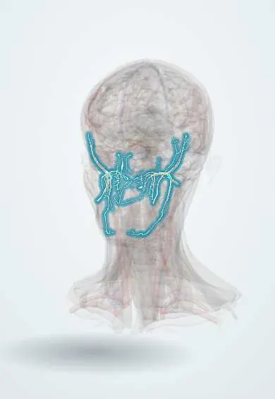
Cardiovascular system · Head and neck
Maxillary artery !
The maxillary artery runs deep behind the jaw to feed the jaws, teeth, chewing muscles, palate and nose; its nasal branch causes back-of-nose bleeds.
Opens in the Durgical app, no sign-up needed.
What it is
A deep external carotid branch behind the jaw with many branches.
What it does
Feeds the jaws, teeth, chewing muscles, palate and nose; its nose branches cause posterior nosebleeds.
Key facts
- Origin
- Terminal branch of the external carotid, behind the mandibular neck
- Branches
- Middle meningeal, inferior alveolar, deep temporal, masseteric, buccal, posterior superior alveolar, infraorbital, descending palatine, sphenopalatine
- Supplies
- Jaws, teeth, muscles of mastication, palate, nose (posterior epistaxis), dura
Relations
- Course
- From the parotid gland behind the neck of the mandible it runs forward between the neck and the sphenomandibular ligament, crosses the lower head of the lateral pterygoid (usually on its surface) and passes through the pterygomaxillary fissure into the pterygopalatine fossa
- Lateral
- Neck and ramus of the mandible and the insertion of the temporalis
- Medial
- Sphenomandibular ligament with the inferior alveolar nerve and lingual nerve; in the fossa, the maxillary nerve and the pterygopalatine ganglion lie close by
How to find it
It cannot be felt: it lies in the infratemporal fossa deep to the ramus of the mandible. Endoscopically, the sphenopalatine artery is found at the sphenopalatine foramen on the lateral nasal wall, behind the back end of the middle turbinate, marked by a small bony crest (crista ethmoidalis).
On imaging
CT angiography shows it leaving the external carotid behind the neck of the mandible, crossing the infratemporal fossa into the pterygopalatine fossa. In posterior epistaxis that does not stop, catheter angiography maps its sphenopalatine branch for embolisation. In juvenile nasopharyngeal angiofibroma, CT and MRI show an enhancing mass widening the pterygopalatine fossa, fed mainly by it.
Clinical notes
- Posterior epistaxis, more common in older people with high blood pressure, runs down the throat and is hard to reach from the front; options include posterior packing, endoscopic sphenopalatine ligation or embolisation
- The inferior alveolar artery runs with the nerve, which is why aspiration is checked before injecting in an inferior alveolar nerve block
- Le Fort I osteotomy and fractures of the condylar neck or the pterygoid plates can tear the artery or its descending palatine branch
Anatomical variants
- It passes superficial to the lateral pterygoid in most people and deep to it in the rest; the proportion differs between populations
Development
Its deep course is borrowed from the embryonic stapedial artery, a second-arch vessel that regresses: the external carotid joins its branches and takes them over, which is why the middle meningeal and inferior alveolar arteries come from the maxillary artery.
Memory aid
Branches of the first part: "DAM I AM" (Deep auricular, Anterior tympanic, Middle meningeal, Inferior alveolar, Accessory Meningeal).
Related conditions
Test yourself
Think of the answer, then tap the question to see it.
How are the three parts of the maxillary artery defined?
By the lateral pterygoid: mandibular (behind the neck of the mandible), pterygoid (on the muscle) and pterygopalatine (in the fossa).
Which branch of the maxillary artery is the main source of posterior epistaxis?
The sphenopalatine artery, entering the nose through the sphenopalatine foramen behind the middle turbinate.
Which two important branches arise from the first part of the maxillary artery?
The middle meningeal artery (to the dura, under the pterion) and the inferior alveolar artery (into the mandibular canal).
Why is aspiration checked before injecting in an inferior alveolar nerve block?
The inferior alveolar artery, a maxillary artery branch, runs with the nerve.
Educational information, not medical advice. For any symptom, see a health professional.
Its names
- Official Latin (TA2)
- arteria maxillaris TA2 4422
- Official Spanish (DeCS)
- Arteria Maxilar DeCS D008438
- English (TA2)
- Maxillary artery
- Also called
- internal maxillary artery · arteria maxillaris interna
Related
Sources: Terminologia Anatomica (TA2); DeCS; Anatomy, Head and Neck, Maxillary Artery; Anatomy of the Human Body (20th ed.): Development of the Vascular System (Lea & Febiger (Gray's Anatomy, 1918), 1918) · adapted, Public domain; Tratado de anatomía humana (9.ª ed.), tomo II, Libro IV, sección 2 Arterias, caps. I-III (sistema de la arteria pulmonar; aorta y ramas del cayado hasta la carótida interna).
Spotted a mistake?We are always improving for the community. Tell us what is wrong and our team will review it.Report a mistake
Representative model, for educational purposes only.