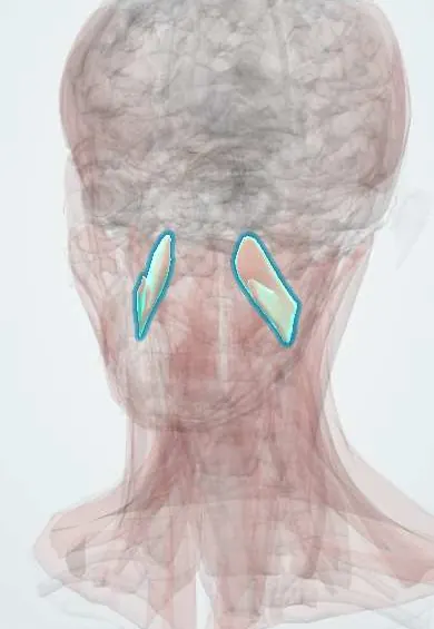
Muscular system · Head and neck
Medial pterygoid !
The medial pterygoid is a chewing muscle on the inner side of the jaw angle, mirroring the masseter; it closes the jaw and helps grind side to side.
Official name: Medial pterygoid muscle
Opens in the Durgical app, no sign-up needed.
What it is
A muscle on the inner side of the jaw angle, mirroring the masseter.
What it does
Closes the jaw and helps grind side to side.
Key facts
- Origin
- Medial surface of the lateral pterygoid plate, maxillary tuberosity
- Insertion
- Medial surface of the ramus and angle of the mandible
- Action
- Elevates and protrudes the mandible; side-to-side grinding
- Nerve
- Nerve to medial pterygoid (V3)
- Blood supply
- Pterygoid branches of the maxillary artery
Relations
- Lateral
- Pterygomandibular space with the lingual nerve, inferior alveolar nerve and vessels and the sphenomandibular ligament, then the ramus of the mandible
- Medial
- Tensor veli palatini, the superior pharyngeal constrictor and the parapharyngeal space
- Above
- Lower head of the lateral pterygoid, which lies between its two heads; the lateral pterygoid plate and maxillary tuberosity at its origin
- Behind
- Deep lobe of the parotid gland
- Below
- Medial surface of the angle of the mandible, near the submandibular gland
How to find it
Felt intraorally with a gloved finger on the inner side of the ramus, behind the last molars, as the person bites; from outside, under the inner side of the angle of the jaw with the head tilted toward that side.
Examination
- Clenching and grinding side to side against resistance; pain on palpation inside the ramus in temporomandibular disorders
- Measuring mouth opening between the incisors to document trismus
On imaging
On axial and coronal MRI or contrast CT it lies on the inner side of the ramus, with the pterygomandibular space between them and the parapharyngeal fat just medial to it. Contrast CT is the usual test for a masticator space abscess after a lower molar infection or a dental block, which causes trismus. MRI shows tumour spread here and denervation atrophy after a mandibular nerve injury.
Clinical notes
- Infection of the pterygomandibular space (often from a lower wisdom tooth) causes trismus with little visible swelling because the muscle lies deep
- Mandibular angle fractures are displaced or held by its pull depending on the direction of the fracture line
- Trismus after radiotherapy for head and neck cancer often involves fibrosis of this muscle
Development
First pharyngeal arch muscle; the shared nerve with the two tensor muscles reflects this common origin.
Related conditions
Test yourself
Think of the answer, then tap the question to see it.
What sling do the medial pterygoid and masseter form?
A sling around the angle of the mandible: masseter outside, medial pterygoid inside; both elevate the jaw.
Why can an inferior alveolar nerve block cause trismus?
The medial pterygoid forms the medial wall of the pterygomandibular space, where the block is given; needle injury to the muscle causes trismus.
Which other muscles share the nerve to medial pterygoid?
The tensor veli palatini and tensor tympani, reflecting their common first-arch origin.
Why does pterygomandibular space infection cause trismus with little visible swelling?
The medial pterygoid lies deep, on the inner side of the ramus.
Educational information, not medical advice. For any symptom, see a health professional.
Its names
- Official Latin (TA2)
- musculus pterygoideus medialis TA2 2113
- Official Spanish (DeCS)
- Músculo pterigoideo medial DeCS D011626
- English (TA2)
- Medial pterygoid muscle
- Also called
- internal pterygoid · musculus pterygoideus internus
Related
Sources: Terminologia Anatomica (TA2); DeCS; Anatomy, Head and Neck, Medial Pterygoid Muscle; Anatomy, Head and Neck, Mastication Muscles.
Spotted a mistake?We are always improving for the community. Tell us what is wrong and our team will review it.Report a mistake
Representative model, for educational purposes only.