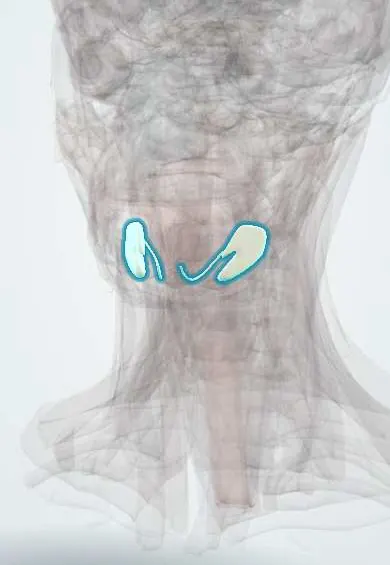
Digestive system · Head and neck
Submandibular gland !
The submandibular gland is a walnut-sized salivary gland under the jaw; it makes most of the resting saliva, and stones often form in its duct.
Opens in the Durgical app, no sign-up needed.
What it is
A walnut-sized salivary gland under the jaw.
What it does
Makes most of the resting saliva; stones often form in its duct.
Key facts
- Where
- Submandibular triangle, wrapping the back edge of the mylohyoid; duct opens beside the frenulum
- Blood supply
- Facial artery
- Nerve
- Parasympathetic: VII (chorda tympani -> submandibular ganglion)
- Function
- Mixed saliva (~70% of resting saliva)
Relations
- Superficial
- Skin, platysma and the investing cervical fascia; the marginal mandibular branch of the facial nerve and the facial vein cross it; submandibular lymph nodes lie on and in its capsule
- Deep
- Mylohyoid in front and hyoglossus behind, with the lingual nerve, submandibular ganglion and hypoglossal nerve on the hyoglossus
- Above
- Submandibular fossa on the inner surface of the mandible, and the medial pterygoid
- Below
- Overlaps the anterior and posterior bellies of the digastric
- Behind
- Stylohyoid and posterior belly of the digastric; the facial artery grooves the back of the gland before hooking over the lower border of the mandible
How to find it
Felt bimanually, with a gloved finger in the floor of the mouth and the other hand under the jaw; a stone may be felt along the duct, and its opening on the sublingual caruncle beside the frenulum shows whether saliva flows.
Examination
- Milking the gland from behind forward while watching the sublingual caruncle: no flow suggests a blocked duct, and pus suggests bacterial sialadenitis
On imaging
Ultrasound shows stones (bright with an acoustic shadow) and a dilated duct; most submandibular stones are radio-opaque and appear on an occlusal radiograph, unlike most parotid stones; CT or sialendoscopy is used when unclear.
Common problems
- Salivary stones block the duct: painful swelling under the jaw at mealtimes.
- Infection behind a stone.
How it is diagnosed
Exam and ultrasound.
Treatment
- Hydration, massage and sour sweets to push small stones out.
- Removing the stone through the mouth or by endoscopy; removing the gland if it keeps happening.
Clinical notes
- Gland excision puts three nerves at risk: marginal mandibular (weak lower lip), lingual (numb tongue and loss of taste on the front two thirds) and hypoglossal (tongue deviates on protrusion)
- Stones near the duct opening can be removed through the floor of the mouth; stones deep in the gland or repeated infection may need sialendoscopy or removal of the gland
- A hard, painless enlargement of the gland in an adult raises suspicion of a tumour, and a larger share of these are malignant than in the parotid
Development
It buds from the lining of the floor of the mouth after the parotid has appeared; the bud grows back and branches into the submandibular region, while the duct keeps its opening beside the frenulum.
Memory aid
The lingual nerve 'double-crosses' Wharton duct: it runs beside it, dips under it and ends up on its inner side.
Related conditions
- Sialolithiasis (salivary stones)
- Acute suppurative sialadenitis (bacterial parotitis)
- Sjögren disease
- IgG4-related disease
Test yourself
Think of the answer, then tap the question to see it.
Why do most salivary stones form in the submandibular gland?
Its saliva is thicker and more alkaline, and the submandibular (Wharton) duct is long and runs uphill.
Around which muscle does the submandibular gland hook?
The free posterior border of the mylohyoid: a large superficial part in the submandibular triangle and a small deep part in the floor of the mouth.
Which three nerves are at risk in submandibular gland excision, and what deficits follow?
Marginal mandibular (weak lower lip), lingual (numb tongue, loss of taste on the front two thirds) and hypoglossal (tongue deviates on protrusion).
Why is the neck incision for the submandibular gland placed about two finger-breadths below the mandible?
To avoid the marginal mandibular branch of the facial nerve, which runs under the platysma over the gland.
Educational information, not medical advice. For any symptom, see a health professional.
Its names
- Official Latin (TA2)
- glandula submandibularis TA2 2810
- Official Spanish (DeCS)
- Glándula Submandibular DeCS D013363
- English (TA2)
- Submandibular gland
- Also called
- submaxillary gland · glandula submaxillaris
Related
Sources: Terminologia Anatomica (TA2); DeCS; Anatomy, Head and Neck, Submandibular Gland; Anatomy, Head and Neck, Salivary Glands; Anatomy of the Human Body (20th ed.): The Digestive Apparatus (Lea & Febiger (Gray's Anatomy, 1918), 1918) · adapted, Public domain.
Spotted a mistake?We are always improving for the community. Tell us what is wrong and our team will review it.Report a mistake
Representative model, for educational purposes only.