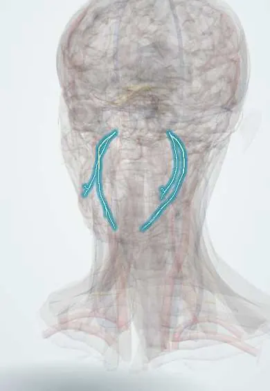
Nervous system · Head and neck
V3 mandibular !
The mandibular nerve (V3), the lowest branch of the trigeminal nerve, gives feeling to the lower face and teeth and powers the chewing muscles.
Official name: Anterior division of mandibular nerve
Opens in the Durgical app, no sign-up needed.
What it is
The lowest trigeminal branch, through the foramen ovale to the jaw.
What it does
Feeling of the lower face and teeth; powers the chewing muscles.
Key facts
- Roots
- V3; foramen ovale
- Moves
- Muscles of mastication, mylohyoid, anterior digastric, tensors; main trunk: nerve to medial pterygoid (and the tensors); anterior trunk: masseteric, deep temporal, lateral pterygoid
- Feeling from
- Lower face, lower teeth, anterior two thirds of the tongue, temple (auriculotemporal); meningeal branch (foramen spinosum); buccal (anterior trunk); auriculotemporal, lingual, inferior alveolar (posterior trunk)
Relations
- Course
- From the trigeminal ganglion, joined by the motor root, straight down through the foramen ovale (it does not enter the cavernous sinus) into the infratemporal fossa, where it divides into its two trunks
- Medial
- The tensor veli palatini, the otic ganglion attached to its inner side, and beyond them the auditory tube
- Lateral
- The lateral pterygoid, which covers it in the infratemporal fossa
- Behind
- The middle meningeal artery on its way to the foramen spinosum; the auriculotemporal nerve splits around it
How to find it
The trunk lies deep to the zygomatic arch and the mandibular notch, the route used for an extraoral mandibular nerve block. Its motor function is felt in the masseter and temporalis when the teeth are clenched.
Examination
- Jaw opening against resistance: deviation toward one side points to weakness of that side's lateral pterygoid (V3)
- Clenching: masseter and temporalis felt for bulk; wasting on one side suggests a lower motor neuron lesion
- Sensation over the chin, lower lip and the skin in front of the ear compared with the other side
On imaging
Contrast MRI, coronal and axial, follows it through the foramen ovale into the masticator space. Perineural spread of a head and neck cancer shows as thickening and enhancement along the nerve, with a widened foramen ovale on CT. Long-standing denervation leaves the chewing muscles on that side small and fatty. CT with bone windows shows the foramen ovale in the skull base.
Clinical notes
- Numb chin syndrome: numbness of the chin and lower lip can be the first sign of cancer spreading to the mandible or the skull base
- Nasopharyngeal and skull base tumours can spread along the nerve through the foramen ovale into the skull
- A mandibular nerve block through the mandibular notch numbs the whole division for surgery or pain procedures
Development
It is the nerve of the first (mandibular) pharyngeal arch and supplies the muscles that come from it: the muscles of mastication, mylohyoid, anterior belly of the digastric, tensor tympani and tensor veli palatini.
Related conditions
Test yourself
Think of the answer, then tap the question to see it.
Through which foramen does the V3 mandibular nerve leave the skull, and does it pass through the cavernous sinus?
The foramen ovale; it drops straight down and does not enter the cavernous sinus.
What are the main branches of the posterior trunk of the V3 mandibular nerve?
The auriculotemporal, lingual and inferior alveolar nerves; the anterior trunk is mainly motor, with the buccal nerve as its one sensory branch.
What is Frey syndrome and which branch of the V3 mandibular nerve is involved?
Gustatory sweating after parotid surgery; the auriculotemporal nerve carries glossopharyngeal parasympathetic fibres from the otic ganglion to the parotid gland.
What can numb chin syndrome be the first sign of?
Cancer spreading to the mandible or the skull base, affecting the V3 mandibular nerve territory of the chin and lower lip.
Educational information, not medical advice. For any symptom, see a health professional.
Its names
- Official Latin (TA2)
- divisio anterior nervi mandibularis TA2 6249
- Official Spanish (DeCS)
- Nervio Mandibular DeCS D008340
- English (TA2)
- Anterior division of mandibular nerve
- Also called
- V3
Related
Sources: Terminologia Anatomica (TA2); DeCS; Anatomy, Head and Neck, Mandibular Nerve; Anatomy of the Human Body (20th ed.): Development of the Muscles (Lea & Febiger (Gray's Anatomy, 1918), 1918) · adapted, Public domain; Anatomy of the Human Body (20th ed.): The Branchial Region (Lea & Febiger (Gray's Anatomy, 1918), 1918) · adapted, Public domain.
Spotted a mistake?We are always improving for the community. Tell us what is wrong and our team will review it.Report a mistake
Representative model, for educational purposes only.