Joints and ligaments · Head and neck
Jaw ligaments !
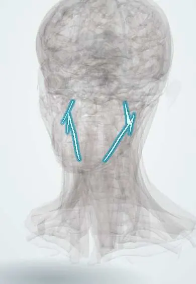
The jaw ligaments are thin bands running from the skull base to the lower jaw and the hyoid bone; they help guide and suspend the jaw.
Official name: Sphenomandibular ligament
Opens in the Durgical app, no sign-up needed.
What it is
Thin bands from the skull base to the jaw and the hyoid.
What it does
Help guide and suspend the jaw and the hyoid.
Key facts
- Attaches
- Sphenomandibular: spine of the sphenoid -> lingula; stylomandibular: styloid -> angle; stylohyoid: styloid -> lesser horn of the hyoid
- Function
- Suspend and guide the mandible and hyoid
- Tight in
- Protrusion (stylomandibular), wide opening (sphenomandibular)
Relations
- Lateral
- Between the sphenomandibular ligament and the ramus pass the maxillary artery and the inferior alveolar nerve and vessels
- Medial
- Medial pterygoid lies medial to the lower sphenomandibular ligament; the pharyngeal wall lies medial to the stylohyoid ligament
- Superficial
- The stylomandibular ligament lies deep to the parotid gland, between it and the submandibular gland
- Deep
- The stylohyoid ligament runs close to the palatine tonsil fossa and the internal carotid artery
How to find it
They are not felt in a normal neck; an elongated styloid process or calcified stylohyoid ligament may be felt as a hard band by a finger in the tonsillar fossa. The lingula, at the end of the sphenomandibular ligament, is the target of the inferior alveolar nerve block.
On imaging
Panoramic radiographs and CT show an elongated styloid process or a calcified stylohyoid ligament; the ligaments themselves are seen only partly on MRI.
Clinical notes
- In the inferior alveolar nerve block the needle aims at the space between the ramus and the sphenomandibular ligament, where the nerve enters the mandibular foramen
- Symptomatic Eagle syndrome can be treated by shortening the styloid process, through the mouth or the neck
Anatomical variants
- An elongated styloid process, or a stylohyoid ligament calcified in part or along its whole length, is a fairly common incidental finding on panoramic radiographs; only a small share of these people have symptoms
Development
The sphenomandibular ligament comes from Meckel cartilage (first arch); the stylohyoid ligament from Reichert cartilage (second arch), which also forms the styloid process and the lesser horn of the hyoid.
Related conditions
Test yourself
Think of the answer, then tap the question to see it.
What is the embryological origin of the sphenomandibular ligament, and where does it end?
A remnant of Meckel cartilage (first arch); it ends at the lingula, beside the inferior alveolar nerve entering the mandibular foramen.
What is Eagle syndrome?
Throat pain on swallowing, sometimes neck pain on turning the head, from a calcified stylohyoid ligament or long styloid process.
Where does the needle aim in an inferior alveolar nerve block, in relation to the jaw ligaments?
The space between the ramus and the sphenomandibular ligament, where the nerve enters the mandibular foramen.
Which ligament is the main stabiliser of the TMJ, rather than the accessory jaw ligaments?
The lateral (temporomandibular) ligament of the capsule.
Educational information, not medical advice. For any symptom, see a health professional.
Its names
- Official Latin (TA2)
- ligamentum sphenomandibulare TA2 1568
- Official Spanish (DeCS)
- Ligamentos esfenomandibular, estilomandibular y estilohioideo
- English (TA2)
- Sphenomandibular ligament
Sources: Terminologia Anatomica (TA2); Anatomy, Head and Neck, Temporomandibular Joint; Anatomy, Head and Neck, Mandibular Foramen.
Spotted a mistake?We are always improving for the community. Tell us what is wrong and our team will review it.Report a mistake
Representative model, for educational purposes only.