Nervous system · Head and neck
Cranial nerve nuclei !
The cranial nerve nuclei are clusters of nerve cells in the brainstem where the cranial nerves start or end; they explain which nerves fail together in strokes.
Official name: Nucleus of oculomotor nerve
Opens in the Durgical app, no sign-up needed.
What it is
Clusters of nerve cells in the brainstem where the cranial nerves start or end.
What it does
Explain which nerves fail together in brainstem strokes.
Key facts
- Where
- Midbrain: III, IV; pons: V, VI, VII, VIII; medulla: IX, X, XII (XI in the upper cervical cord)
- Function
- Motor nuclei lie near the midline, sensory ones laterally; brainstem strokes combine a cranial nerve sign with opposite-side limb signs
Relations
- Behind
- Many lie under the floor of the fourth ventricle: the facial colliculus (abducens nucleus), the hypoglossal and vagal trigones and the vestibular area
- In front
- Descending corticospinal fibres in the base of the brainstem, which is why lesions combine cranial nerve and limb signs
- Contents
- Midbrain: oculomotor, Edinger–Westphal, trochlear; pons: trigeminal motor and main sensory, abducens, facial, salivatory, vestibular and cochlear; medulla: ambiguus, solitary, dorsal vagal, hypoglossal
Examination
- Pupils and eye movements: oculomotor, trochlear and abducens nuclei, linked by the medial longitudinal fasciculus
- Corneal reflex and facial movement: trigeminal and facial nuclei in the pons
- Gag reflex, palate elevation and voice: nucleus ambiguus and the glossopharyngeal and vagus nerves
- Tongue protrusion: deviation toward the weak side in a hypoglossal nucleus or nerve lesion
On imaging
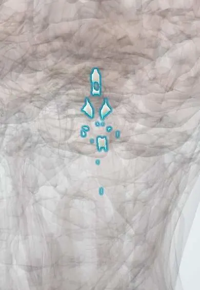
The nuclei are not seen one by one on routine imaging. MRI (axial T2, FLAIR, DWI) finds a brainstem lesion and the nucleus is inferred from its level: midbrain for III and IV, pons for V to VIII, medulla for IX to XII. On the floor of the fourth ventricle, the facial colliculus marks the abducens nucleus. CT shows the brainstem poorly.
Clinical notes
- Matching the cranial nerve sign to its nucleus gives the level: oculomotor (Weber, midbrain), abducens and facial (Millard–Gubler, pons), hypoglossal (medial medulla), ambiguus (lateral medulla)
- A lesion of the spinal trigeminal nucleus in the lateral medulla removes pain and temperature from the same side of the face
- The four vestibular nuclei (superior, lateral or Deiters, medial, inferior) lie under the vestibular area; lateral medullary (Wallenberg) infarcts hit them: vertigo, nystagmus and falling toward the lesion
Development
As in the spinal cord, a groove (sulcus limitans) separates the motor basal plate from the sensory alar plate. When the fourth ventricle opens, the plates fold outward, so in its floor motor nuclei lie medial and sensory nuclei lateral.
Memory aid
Rule of 4: four cranial nerves above the pons (I–IV), four in the pons (V–VIII), four in the medulla (IX–XII); the four motor nerves that divide evenly into 12 (III, IV, VI, XII) have nuclei near the midline.
Related conditions
Test yourself
Think of the answer, then tap the question to see it.
Which cranial nerve nuclei lie in the midbrain, pons and medulla?
Midbrain: III, IV; pons: V, VI, VII, VIII; medulla: IX, X, XII (XI in the upper cervical cord).
What does a crossed brainstem syndrome consist of?
A cranial nerve sign on the side of the lesion plus limb signs on the other side, as the descending corticospinal fibres run in front of the nuclei.
Which nuclei handle swallowing and voice, and taste and visceral sensation?
Nucleus ambiguus (glossopharyngeal, vagus): swallowing and voice; nucleus of the solitary tract: taste and visceral sensation.
Why does an upper motor neuron lesion spare the forehead but a facial nucleus lesion does not?
The lower face part of the facial motor nucleus gets only crossed cortical input, so an upper motor neuron lesion spares the forehead; a nuclear or nerve lesion does not.
Educational information, not medical advice. For any symptom, see a health professional.
Its names
- Official Latin (TA2)
- nucleus nervi oculomotorius TA2 5892
- Official Spanish (DeCS)
- Núcleos de los nervios craneales
- English (TA2)
- Nucleus of oculomotor nerve
- Also called
- oculomotor nucleus · nucleus oculomotorius · nucleus of cranial nerve III
Sources: Terminologia Anatomica (TA2); Neuroanatomy, Cranial Nerve; Neuroanatomy, Brainstem; 13.2 The Central Nervous System (OpenStax (Rice University), 2022) · adapted, CC BY 4.0; Anatomía humana (4.ª ed.), tomo 1, cap. 52: Vías vestibulares.
Spotted a mistake?We are always improving for the community. Tell us what is wrong and our team will review it.Report a mistake
Representative model, for educational purposes only.