Benign paroxysmal positional vertigo (BPPV) !
Benign paroxysmal positional vertigo (BPPV) is brief spinning dizziness when the head moves, caused by loose crystals in the inner ear.
Updated 2026-10-29
Structures involved
Tap one to see what it is and open it in 3D.
- Semicircular canals
- Vestibule
- Vestibulocochlear nerve (VIII)
- Temporal bones
- Cerebellum
- Cranial nerve nuclei
In histology
In the clinical exam
In physiology
What it is
Brief spells of spinning vertigo triggered by changes in head position, caused by loose otoconia (calcium carbonate crystals) in a semicircular canal. It is the most common cause of peripheral vertigo.
Who gets it
Most common in middle-aged and older adults, more often in women. Many cases have no clear cause; it often recurs.
Causes
- Idiopathic (most cases)
- Head trauma
- After vestibular neuritis or other inner ear disease
- Prolonged bed rest or head positioning (e.g. dental or surgical procedures)
Risk factors
- Older age
- Female sex
- Head injury
- Previous inner ear disease (vestibular neuritis, Ménière disease)
- Osteoporosis and low vitamin D (association reported)
- Migraine
Pathophysiology
Otoconia detach from the utricle and drift into a semicircular canal, most often the posterior one because it is the lowest when lying down. When the head moves in the plane of that canal, the particles move with gravity and drag the endolymph, deflecting the cupula and causing vertigo with a typical nystagmus until they settle.
Classification
- Posterior canal (most common)
- Horizontal (lateral) canal
- Anterior canal (rare)
- Canalithiasis (free particles) vs cupulolithiasis (particles stuck to the cupula)
Symptoms and signs
- Spinning vertigo lasting less than about a minute
- Triggered by rolling over in bed, lying down, looking up or bending forward
- Nausea, sometimes vomiting
- Imbalance between spells
- No hearing loss or other neurological symptoms
Diagnosis
- Clinical
- Positional tests: the Dix–Hallpike manoeuvre shows upbeating torsional nystagmus with a short delay, lasting under a minute, in posterior canal BPPV. The supine roll test shows horizontal nystagmus in horizontal canal BPPV.
- Tests
- None routinely. Audiometry if hearing symptoms; vestibular function testing in atypical cases.
- Imaging
- Not needed when positional tests are typical. MRI of the brain if signs are atypical, neurological findings are present or treatment fails.
Differential diagnosis
- Vestibular neuritis
- Ménière disease
- Vestibular migraine
- Posterior circulation stroke or TIA
- Orthostatic hypotension
- Central positional nystagmus (cerebellar lesion)
Goal of treatment
Move the otoconia out of the affected canal to stop the vertigo, and reduce falls and recurrences.
Treatment
- Procedure
- Canalith repositioning manoeuvres: the Epley manoeuvre for posterior canal BPPV; roll manoeuvres (barbecue roll, Gufoni) for the horizontal canal. Often effective after one or a few sessions.
- Rehabilitation
- Vestibular rehabilitation for residual imbalance and in older adults at risk of falls; some patients learn self-repositioning manoeuvres.
- Medical
- Vestibular suppressants (antihistamines, benzodiazepines) only briefly for severe nausea; they are not a treatment for BPPV and can delay compensation.
- Surgical
- Surgical occlusion of the posterior canal in rare, persistent cases that fail repeated manoeuvres.
Complications
- Falls and fractures, especially in older adults
- Recurrence
- Anxiety and avoidance of movement
- Persistent dizziness after successful repositioning
Prognosis
Good: most cases resolve with repositioning, and many resolve on their own. Recurrence is common over the following years.
Test yourself
Think of the answer, then tap the question to see it.
Which semicircular canal is most often affected in BPPV?
The posterior canal, because it is the lowest part of the labyrinth when lying down.
Which test confirms posterior canal BPPV?
The Dix–Hallpike manoeuvre, showing delayed, brief upbeating torsional nystagmus.
What is the treatment of choice for posterior canal BPPV?
A canalith repositioning manoeuvre such as the Epley manoeuvre.
Which features of positional vertigo suggest a central cause rather than BPPV?
Purely vertical or direction-changing nystagmus without delay or fatigue, and other neurological signs such as ataxia or diplopia.
Related conditions
- Ménière disease
- Vestibular schwannoma (acoustic neuroma)
- Ischaemic stroke
- Migraine
- Transient ischaemic attack
Educational information, not medical advice. For any symptom, see a health professional.
Sources: Benign Paroxysmal Positional Vertigo (StatPearls (NCBI Bookshelf), 2025).
Spotted a mistake?We are always improving for the community. Tell us what is wrong and our team will review it.Report a mistake
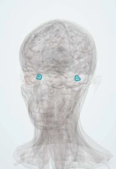
Representative model, for educational purposes only.