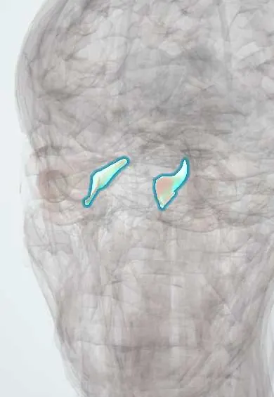
Medial rectus !
The medial rectus is one of the six muscles that move the eyeball, on the nose side of the eye; it turns the eye toward the nose.
Official name: Medial rectus muscle
Opens in the Durgical app, no sign-up needed.
What it is
One of the six muscles that move the eyeball, inside the eye socket.
What it does
Turns the eye toward the nose.
Key facts
- Origin
- Common tendinous ring (annulus of Zinn)
- Insertion
- Sclera medially, ~5.5 mm behind the limbus
- Action
- Adduction
- Nerve
- Oculomotor nerve (III), inferior division
- Blood supply
- Ophthalmic artery (muscular branches)
Relations
- Medial
- Thin medial orbital wall (lamina papyracea of the ethmoid bone), with the ethmoid cells behind it
- Above
- Superior oblique; between the two run the ophthalmic artery, the nasociliary nerve and their ethmoidal branches
- Lateral
- Globe and optic nerve; the inferior division of the oculomotor nerve enters its lateral (ocular) surface
How to find it
Its tendon inserts closest to the cornea, about 5.5 mm from the medial limbus, so it is the easiest rectus to reach through a nasal conjunctival incision. Further back it runs along the medial wall next to the paper-thin lamina papyracea of the ethmoid, with the superior oblique above it.
Examination
- H-pattern: the person looks straight in toward the nose; weak adduction points to the medial rectus or the oculomotor nerve (III)
- Convergence: a target brought toward the nose; adduction that fails in side gaze but works in convergence places the lesion in the medial longitudinal fasciculus
- Cover–uncover and alternate cover tests: an eye that moves out to take up fixation was turned in (esotropia); one that moves in was turned out (exotropia)
On imaging
Coronal CT or MRI of the orbits shows enlargement of the belly in thyroid eye disease and crowding of the optic nerve by enlarged medial recti at the apex; CT shows the lamina papyracea in sinus disease and medial wall fractures.
Common problems
- Sixth-nerve palsy: the eye cannot turn out, causing side-by-side double vision.
- Strabismus (squint) when the eye muscles are unbalanced.
How it is diagnosed
Eye movement and alignment tests; scans if a nerve palsy is new.
Treatment
- Prisms or patching; botulinum toxin; eye muscle surgery to rebalance the muscles.
Clinical notes
- Strabismus in children: infantile esotropia appears in the first months; accommodative esotropia appears later in long-sighted children and improves with glasses. Surgery often weakens or tightens the horizontal recti
- An untreated squint or unequal focus in early childhood causes amblyopia: the brain suppresses one eye and its vision fails to develop; treatment works only while the visual system is still maturing
- Endoscopic sinus surgery can breach the lamina papyracea and injure the medial rectus; ethmoid sinusitis spreads to the orbit by the same route
- A medial wall fracture can trap the muscle and limit abduction
Development
Forms from head mesoderm around the optic stalk, within the common mass that gives rise to all the muscles of the oculomotor nerve.
Related conditions
Test yourself
Think of the answer, then tap the question to see it.
What is the action and nerve supply of the medial rectus?
Pure adduction; supplied by the inferior division of the oculomotor nerve (III).
What is internuclear ophthalmoplegia, and what does it suggest in a young adult?
A medial longitudinal fasciculus lesion: the medial rectus fails in side gaze but works in convergence; in a young adult it suggests multiple sclerosis.
Why can endoscopic sinus surgery injure the medial rectus?
It lies against the thin lamina papyracea of the ethmoid; breaching it reaches the muscle, and ethmoid sinusitis spreads to the orbit the same way.
What happens to an eye with an untreated squint in early childhood?
Amblyopia: the brain suppresses that eye and its vision fails to develop; treatment works only while the visual system is maturing.
Educational information, not medical advice. For any symptom, see a health professional.
Its names
- Official Latin (TA2)
- musculus rectus medialis TA2 2044
- Official Spanish (DeCS)
- Músculo recto medial DeCS D009801
- English (TA2)
- Medial rectus muscle
- Also called
- internal rectus · musculus rectus nasalis bulbi
Related
Sources: Terminologia Anatomica (TA2); DeCS; Anatomy, Head and Neck: Eye Medial Rectus Muscles; Anatomy, Head and Neck, Eye Extraocular Muscles; Anatomy of the Human Body (20th ed.): The Accessory Organs of the Eye (Lea & Febiger (Gray's Anatomy, 1918), 1918) · adapted, Public domain; Anatomy of the Human Body (20th ed.): Development of the Muscles (Lea & Febiger (Gray's Anatomy, 1918), 1918) · adapted, Public domain.
Spotted a mistake?We are always improving for the community. Tell us what is wrong and our team will review it.Report a mistake
Representative model, for educational purposes only.