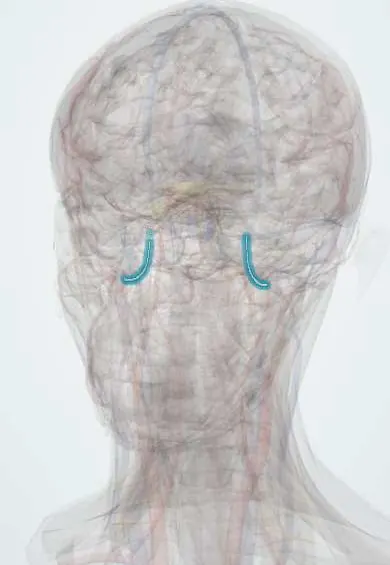
Cardiovascular system · Head and neck
Middle meningeal artery !
The middle meningeal artery runs on the inside of the skull at the temple; a skull fracture there can tear it and cause an epidural haematoma.
Opens in the Durgical app, no sign-up needed.
What it is
An artery under the pterion, inside the skull; a fracture there can tear it and cause an epidural haematoma.
What it does
Feeds the dura and skull; a skull fracture can tear it, causing an epidural haematoma.
Key facts
- Origin
- Maxillary artery; enters through the foramen spinosum
- Branches
- Frontal and parietal branches
- Supplies
- Dura and skull; frontal branch under the pterion (epidural haematoma)
Relations
- Course
- From the first part of the maxillary artery in the infratemporal fossa it rises deep to the lateral pterygoid, through the loop of the auriculotemporal nerve, enters the middle cranial fossa through the foramen spinosum and runs out across its floor; the frontal branch climbs to the pterion and the parietal branch runs backward
- Superficial
- Inner table of the skull, which it grooves; at the pterion, where the frontal, parietal, temporal and sphenoid bones meet, the bone is thin
- Deep
- Dura mater, in whose outer layer it runs, and beneath it the temporal and frontal lobes
How to find it
The pterion, over the frontal branch, lies about two finger-breadths above the zygomatic arch and a thumb's breadth behind the frontal process of the zygomatic bone.
On imaging
Non-contrast CT: a bright biconvex collection under the temple, often with a fracture crossing the vessel groove on bone windows; darker swirls inside it suggest active bleeding. On plain skull films the vascular grooves can look like a fracture.
Common problems
- Epidural haematoma: bleeding between skull and dura, usually from the middle meningeal artery after a blow; the person may seem fine at first, then worsens.
- Subdural haematoma: bleeding under the dura from torn veins, common in older adults and people on blood thinners.
How it is diagnosed
Emergency CT of the head.
Treatment
- Observation for small bleeds.
- Urgent craniotomy or burr holes to remove the blood.
Clinical notes
- Arterial bleeding grows fast: the clot can push the medial temporal lobe through the tentorial notch (uncal herniation), compressing the oculomotor nerve and the brainstem
- A large epidural haematoma is an emergency treated by surgical evacuation through a craniotomy
- Embolisation of the middle meningeal artery is used to lower the recurrence of chronic subdural haematoma, whose membranes it supplies
- When cut it bleeds from both ends, because its branches anastomose with the other meningeal arteries and with the opposite side; at the pterion its frontal branch may run in a bony tunnel, so a fracture there tears it easily
Anatomical variants
- Rarely the ophthalmic artery arises from it, or the two share a large orbital link; this matters before embolisation
- An accessory meningeal artery may arise from it or from the maxillary artery
Development
It is a remnant of the embryonic stapedial artery, whose trunk passes between the two roots of the auriculotemporal nerve; its small orbital twigs are what is left of the stapedial branch to the orbit, explaining links with the ophthalmic artery.
Memory aid
Epidural = lens, stopped by the sutures; subdural = crescent, crossing the sutures.
Related conditions
Test yourself
Think of the answer, then tap the question to see it.
From which artery does the middle meningeal artery arise, and through which foramen does it enter the skull?
The first part of the maxillary artery; it enters the middle cranial fossa through the foramen spinosum.
What is the classic course of an epidural haematoma from a torn middle meningeal artery?
A blow to the temple tears the frontal branch under the pterion; a lucid interval, then drowsiness, a dilated pupil on that side and weakness of the other side.
What does an epidural haematoma look like on CT, and why?
Lens-shaped (biconvex) and stopped by the sutures, where the dura is firmly attached to the bone.
Why is the middle meningeal artery embolised in chronic subdural haematoma?
It supplies the haematoma's membranes; embolisation lowers the recurrence.
Educational information, not medical advice. For any symptom, see a health professional.
Its names
- Official Latin (TA2)
- arteria meningea media TA2 4431
- Official Spanish (DeCS)
- Arteria meníngea media DeCS D008576
- English (TA2)
- Middle meningeal artery
- Also called
- arteria meningica media
Related
Sources: Terminologia Anatomica (TA2); DeCS; Neuroanatomy, Middle Meningeal Arteries; Anatomy of the Human Body (20th ed.): Development of the Vascular System (Lea & Febiger (Gray's Anatomy, 1918), 1918) · adapted, Public domain; Tratado de anatomía humana (9.ª ed.), tomo II, Libro IV, sección 2 Arterias, caps. I-III (sistema de la arteria pulmonar; aorta y ramas del cayado hasta la carótida interna).
Spotted a mistake?We are always improving for the community. Tell us what is wrong and our team will review it.Report a mistake
Representative model, for educational purposes only.