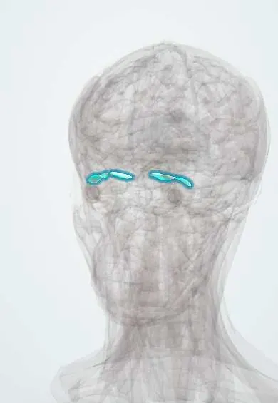
Superior oblique !
The superior oblique is an eye-moving muscle whose tendon runs through a pulley in the eye socket; it turns the eye down and in and is supplied by nerve IV.
Official name: Superior oblique muscle
Opens in the Durgical app, no sign-up needed.
What it is
One of the six muscles that move the eyeball, inside the eye socket.
What it does
Turns the eye down and in and rotates it inward; its tendon runs through a pulley (trochlea); supplied by nerve IV.
Key facts
- Origin
- Body of the sphenoid above the optic canal
- Insertion
- Posterolateral sclera behind the equator, after passing through the trochlea
- Action
- Intorsion; depression (best in adduction); abduction
- Nerve
- Trochlear nerve (IV)
- Blood supply
- Ophthalmic artery (muscular branches)
Relations
- Course
- From the body of the sphenoid it runs forward along the upper medial orbit to the trochlea at the front of the roof; the tendon turns back and outward, passes under the superior rectus and inserts on the upper outer back of the globe
- Above
- Orbital roof (frontal bone); the trochlear nerve enters its upper surface
- Below
- Medial rectus, with the ophthalmic artery and nasociliary nerve between them
- Lateral
- Levator palpebrae superioris and superior rectus
- Medial
- Medial orbital wall (frontal and ethmoid bones)
How to find it
The trochlea sits just inside the upper inner orbital rim, a few millimetres behind it; it can be felt as a small firm point under the rim and is tender in trochleitis. Surgery at this corner (frontal sinus, upper medial lid) can damage it.
Examination
- H-pattern: the person looks in toward the nose, then down; poor depression in adduction isolates the superior oblique
- Parks–Bielschowsky three-step test: a hypertropia that worsens on looking to the opposite side and on tilting the head toward the same side identifies a weak superior oblique
On imaging
Coronal CT or MRI shows its belly in the upper medial orbit, above the medial rectus. On CT the trochlea at the upper inner rim often calcifies with age, a normal finding. In a congenital fourth nerve palsy MRI may show a small or absent muscle belly; in an acquired palsy, MRI of the brainstem looks for the cause. In trochleitis, MRI shows inflammation around the trochlea.
Common problems
- Sixth-nerve palsy: the eye cannot turn out, causing side-by-side double vision.
- Strabismus (squint) when the eye muscles are unbalanced.
How it is diagnosed
Eye movement and alignment tests; scans if a nerve palsy is new.
Treatment
- Prisms or patching; botulinum toxin; eye muscle surgery to rebalance the muscles.
Clinical notes
- Head trauma is the commonest acquired cause of a fourth-nerve palsy; congenital palsies often show a long-standing head tilt in old photographs
- Brown syndrome: the tendon cannot slide through the trochlea, so the eye fails to rise in adduction; congenital, or after inflammation or surgery
- In an oculomotor (III) palsy the superior oblique still works: on attempted downgaze the eye intorts, showing that the trochlear nerve is intact
Development
Forms from head mesoderm near the optic stalk, apart from the oculomotor mass; congenital fourth-nerve palsy, often with a lax, long tendon, is a frequent cause of a vertical squint present since early childhood.
Memory aid
"LR6 SO4, rest 3": superior oblique → trochlear (IV); lateral rectus → abducens (VI); all the others → oculomotor (III).
Related conditions
Test yourself
Think of the answer, then tap the question to see it.
What are the actions of the superior oblique, and why?
Intorsion, depression and abduction: its tendon turns at the trochlea, so it pulls the back of the eye from the front.
In which gaze direction is the superior oblique tested?
Looking down and in: with the eye adducted it is the main depressor.
What are the signs of a fourth-nerve palsy?
Vertical double vision worse looking down and in, the affected eye higher, and the head tilted to the opposite side.
What is unique about the trochlear nerve that supplies the superior oblique?
It is the thinnest cranial nerve, the only one leaving the back of the brainstem and crossing over, with the longest intracranial course.
What is Brown syndrome?
The superior oblique tendon cannot slide through the trochlea, so the eye fails to rise in adduction.
Educational information, not medical advice. For any symptom, see a health professional.
Its names
- Official Latin (TA2)
- musculus obliquus superior bulbi oculi TA2 2048
- Official Spanish (DeCS)
- Músculo oblicuo superior DeCS D009801
- English (TA2)
- Superior oblique muscle
- Also called
- musculus obliquus superior bulbi · Oblicuo mayor
Related
Sources: Terminologia Anatomica (TA2); DeCS; Anatomy, Head and Neck: Eye Superior Oblique Muscle; Anatomy, Head and Neck, Eye Extraocular Muscles; Anatomy of the Human Body (20th ed.): Development of the Muscles (Lea & Febiger (Gray's Anatomy, 1918), 1918) · adapted, Public domain.
Spotted a mistake?We are always improving for the community. Tell us what is wrong and our team will review it.Report a mistake
Representative model, for educational purposes only.