Skeletal system · Head and neck
Sphenoid sinus !
The sphenoid sinus is an air space deep in the skull base, just under the pituitary gland; surgeons pass through it to reach the pituitary from the nose.
Official name: Sinus of sphenoid bone
Opens in the Durgical app, no sign-up needed.
What it is
An air space in the sphenoid body, just under the pituitary.
What it does
Surgeons pass through it to reach the pituitary from the nose.
Key facts
- Where
- Body of the sphenoid, below the sella; drains to the sphenoethmoidal recess
- Blood supply
- Posterior ethmoidal, sphenopalatine arteries
- Nerve
- V1, V2
- Function
- Surgical route to the pituitary
Relations
- Above
- Sella turcica with the pituitary gland; the optic chiasm above and in front
- Lateral
- Cavernous sinus with the internal carotid artery and nerves III, IV, V1, V2 and VI; the optic canal above and lateral
- In front
- Posterior ethmoid cells and the sphenoethmoidal recess of the nasal cavity
- Below
- Roof of the nasopharynx; the pterygoid canal with its nerve in the floor
- Behind
- Clivus, with the basilar artery and pons beyond
- Medial
- Intersinus septum, often off the midline and sometimes attached over the carotid canal
How to find it
It cannot be felt. Endoscopically its ostium is found in the sphenoethmoidal recess, medial to the superior concha, close to the back of the septum.
On imaging
CT shows its pneumatisation, its septa and where they attach, and gaps in the bone over the optic nerve or internal carotid artery before surgery; MRI shows the sella, the cavernous sinus and complications of sphenoid sinusitis.
Common problems
- Sinusitis: blocked nose, thick discharge, pressure in the cheeks or teeth.
- Most acute sinusitis is viral; some becomes chronic.
How it is diagnosed
Symptoms and exam; nasal endoscopy and CT for chronic or complicated cases.
Treatment
- Saline rinses, nasal steroid sprays, pain relief; antibiotics only for bacterial cases.
- Endoscopic sinus surgery for chronic sinusitis.
Clinical notes
- A septum attached over the internal carotid artery can tear the artery if twisted off during surgery
- Sphenoid sinusitis or a mucocele can compress the optic nerve, with visual loss
- CSF leak after transsphenoidal surgery is a recognised complication, because the floor of the sella is opened
Anatomical variants
- Pneumatisation types by relation to the sella: conchal (minimal, mostly children), presellar and sellar (most adults); the sellar type gives the widest surgical access
- Lateral recesses into the greater wings or pterygoid processes, and pneumatised anterior clinoid processes
Development
Tiny at birth; pneumatisation of the sphenoid body begins in early childhood and extends back under the sella through later childhood and adolescence.
Related conditions
Test yourself
Think of the answer, then tap the question to see it.
Which surgical route passes through the sphenoid sinus?
Transsphenoidal surgery to the pituitary gland: through the nose, the sphenoid sinus and the floor of the sella turcica.
Which structures bulge into the lateral wall of the sphenoid sinus, and why is that dangerous?
The internal carotid artery and the optic nerve; the bone over them can be very thin or absent.
Why is isolated sphenoid sinusitis serious?
It causes deep headache and can spread to the cavernous sinus or optic nerve, causing visual loss and eye movement palsies.
Where does the sphenoid sinus drain?
Into the sphenoethmoidal recess, medial to the superior concha.
Educational information, not medical advice. For any symptom, see a health professional.
Its names
- Official Latin (TA2)
- sinus ossis sphenoidei TA2 600
- Official Spanish (DeCS)
- Seno Esfenoidal DeCS D013101
- English (TA2)
- Sinus of sphenoid bone
- Also called
- bony sphenoidal sinus · sinus sphenoideus · sinus sphenoidalis
Related
Sources: Terminologia Anatomica (TA2); DeCS; Anatomy, Head and Neck, Nose Paranasal Sinuses; Anatomy, Head and Neck, Nose Sinuses.
Spotted a mistake?We are always improving for the community. Tell us what is wrong and our team will review it.Report a mistake
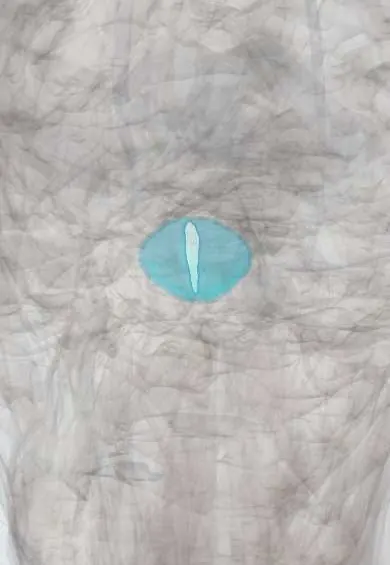
Representative model, for educational purposes only.