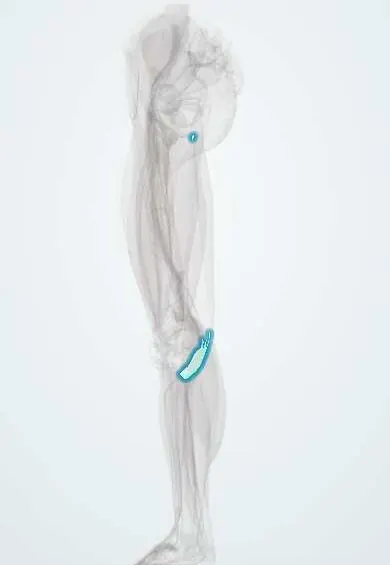
Semimembranosus tendon !
The semimembranosus tendon is the hamstring tendon that wraps the back inner corner of the tibia and tightens the back of the knee capsule.
Official name: Tendon of semimembranosus
Opens in the Durgical app, no sign-up needed.
What it is
The broad hamstring tendon wrapping the back inner corner of the tibia.
What it does
Helps bend the knee and turn the shin inward; supports the knee from the inner side.
Key facts
- Attaches
- Main insertion in the posterior groove of the medial tibial condyle; expansions to the oblique popliteal ligament, posterior capsule, medial meniscus and popliteus fascia
- Function
- Knee flexion and medial rotation; hip extension; stabilizes the posteromedial corner; the semimembranosus–gastrocnemius bursa between it and the medial gastrocnemius is where a Baker cyst forms
- Tight in
- Hip flexion with the knee extended
Relations
- Superficial
- Semitendinosus tendon, which lies on it
- Lateral
- Medial head of gastrocnemius, with the semimembranosus–gastrocnemius bursa between them; further out, the popliteal artery, popliteal vein and tibial nerve
- Deep
- Posterior knee joint capsule and the oblique popliteal ligament
- Medial
- Superficial MCL, under which its anterior (tibial) arm passes
How to find it
Prone with the knee bent against resistance, it is felt as the broad tendon on either side of and deep to the semitendinosus cord; its insertion is felt just below the joint line on the back of the medial tibial condyle.
On imaging
MRI shows its arms around the posteromedial corner, and insertional tendinopathy as thickening, high signal or bone oedema; axial images show a Baker's cyst between it and the medial gastrocnemius tendon.
Common problems
- Strains or tendinitis, mostly in sports with sprinting or kicking.
How it is diagnosed
Physical exam; ultrasound or MRI if needed.
Treatment
- Rest, ice and progressive physical therapy.
Clinical notes
- Posteromedial corner injuries often accompany ACL tears; ramp lesions of the medial meniscus lie close to where its capsular arm attaches
- A separate semimembranosus bursa, between the tendon and the tibia, can also swell and mimic a Baker's cyst
Related conditions
Test yourself
Think of the answer, then tap the question to see it.
Where is the main insertion of the semimembranosus tendon?
In the posterior groove of the medial tibial condyle, with expansions to the oblique popliteal ligament, posterior capsule and medial meniscus.
Through which gap does a Baker's cyst communicate with the knee joint?
Between the semimembranosus tendon and the tendon of the medial head of gastrocnemius.
What does the semimembranosus do to the medial meniscus as the knee bends?
Its arms tension the posteromedial capsule and pull the medial meniscus backward.
Educational information, not medical advice. For any symptom, see a health professional.
Its names
- Official Latin (TA2)
- musculus semimembranosus TA2 2642
- Official Spanish (DeCS)
- Tendón del semimembranoso DeCS D013710
- English (TA2)
- Tendon of semimembranosus
- Also called
- musculus semimembranaceus
Related
Sources: Terminologia Anatomica (TA2); DeCS; Anatomy, Bony Pelvis and Lower Limb, Hamstring Muscle; Anatomy, Bony Pelvis and Lower Limb: Posterior Thigh Muscles.
Spotted a mistake?We are always improving for the community. Tell us what is wrong and our team will review it.Report a mistake
Representative model, for educational purposes only.