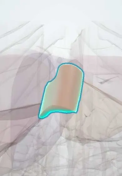
Liver segment I !
Liver segment I (caudate lobe) is a small part at the back of the liver, beside the vena cava; it drains on its own and often enlarges in liver disease.
Official name: Posterior segment of liver
Opens in the Durgical app, no sign-up needed.
What it is
Liver segment I: the caudate lobe, at the back next to the vena cava, with its own drainage.
What it does
Because each segment has its own supply, surgeons can remove one or more segments and leave the rest working.
Key facts
- Where
- Caudate lobe: posterior, between IVC, ligamentum venosum and porta hepatis
- Blood supply
- Branches of both right and left portal vein and hepatic artery; drains by short hepatic veins straight into IVC
- Nerve
- Hepatic plexus (celiac sympathetic and vagal fibres) along its portal pedicle, shared with the whole liver
- Function
- Independent drainage often spares it in hepatic vein obstruction (Budd–Chiari); bile drains to both hepatic ducts
Relations
- In front
- Porta hepatis, hilar plate and portal bifurcation, which separate it from liver segment IV
- Behind
- Inferior vena cava, which it partly wraps; a fibrous band (the caval ligament) may join it to liver segment VII behind the cava
- Below
- Papillary and caudate processes pointing toward the porta hepatis and the epiploic foramen; the lobe bulges into the superior recess of the lesser sac, with the lesser omentum on its left
How to find it
Hidden from the front: it lies on the back of the liver between the inferior vena cava and the fissure for the ligamentum venosum, reached in surgery through the lesser omentum or by turning the liver over.
On imaging
On axial CT or MRI it sits between the IVC behind and the fissure for the ligamentum venosum in front. In cirrhosis, a caudate-to-right-lobe width ratio above about 0.65 is a classic sign; in Budd–Chiari it may be the only part that enhances normally.
Common problems
- Fatty liver disease, very common with obesity and diabetes.
- Hepatitis (viral, alcohol).
- Cirrhosis: scarring that can lead to liver failure and portal hypertension.
- Liver cancer and metastases.
How it is diagnosed
Blood tests (liver enzymes), ultrasound, elastography, CT or MRI, biopsy.
Treatment
- Treat the cause (weight loss, antiviral medicines, stopping alcohol); surgery to remove segments; liver transplant for failure.
Clinical notes
- Isolated caudate resection is among the hardest liver operations: the lobe is deep, and its short hepatic veins enter the inferior vena cava directly and must be tied one by one
- Hilar cholangiocarcinoma often spreads into the caudate bile ducts, so the caudate lobe is usually removed as part of its resection
- Caudate tumours pressing on the inferior vena cava may need total vascular exclusion: the hepatic pedicle and the cava above and below the liver are clamped during the resection
Anatomical variants
- A large papillary process can lie separate from the rest of the lobe and mimic a node near the pancreatic head on CT
Development
The fissure that marks its front edge holds the ligamentum venosum, the remnant of the fetal ductus venosus that carried umbilical blood past the liver to the IVC.
Test yourself
Think of the answer, then tap the question to see it.
Why is liver segment I (caudate lobe) often spared in Budd–Chiari syndrome?
Its short hepatic veins drain straight into the IVC, so it enlarges while the rest of the liver shrinks.
What is the blood supply of liver segment I?
Branches of both the right and left portal vein and hepatic artery; it is neither right nor left liver.
Why is liver segment I usually removed with a hilar cholangiocarcinoma?
The tumour often spreads into the caudate bile ducts, which drain to both hepatic ducts.
Educational information, not medical advice. For any symptom, see a health professional.
Its names
- Official Latin (TA2)
- segmentum posterius hepatis TA2 3073
- Official Spanish (DeCS)
- Segmento I del hígado DeCS D008099
- English (TA2)
- Posterior segment of liver
- Also called
- segment I of liver · lobus caudatus · caudate lobe
Sources: Terminologia Anatomica (TA2); DeCS; Diagnostic Imaging of the Liver; Anatomy, Abdomen and Pelvis: Liver; 23.6 Accessory Organs in Digestion: The Liver, Pancreas, and Gallbladder (OpenStax (Rice University), 2022) · adapted, CC BY 4.0.
Spotted a mistake?We are always improving for the community. Tell us what is wrong and our team will review it.Report a mistake
Representative model, for educational purposes only.