Joints and ligaments · Lower limb
Sacrospinous ligament !
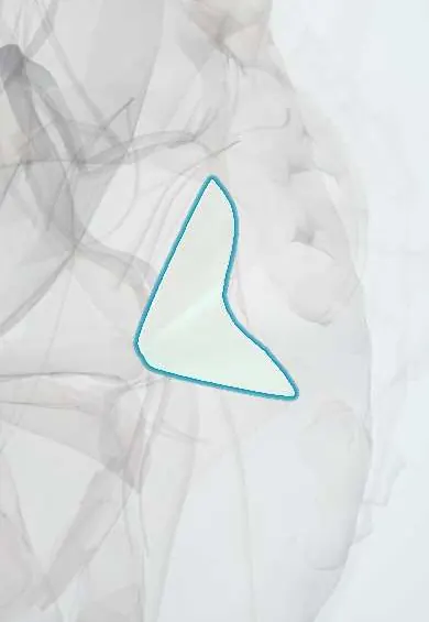
The sacrospinous ligament is a short band from the sacrum to a bony spine on the hip bone; it steadies the pelvis.
Opens in the Durgical app, no sign-up needed.
What it is
A short, triangular band from the sacrum and coccyx to the ischial spine, a point on the hip bone.
What it does
Steadies the sacrum and helps form the openings through which the sciatic nerve and other nerves leave the pelvis.
Key facts
- Attaches
- Lateral sacrum and coccyx to ischial spine
- Function
- Resists nutation; separates greater from lesser sciatic foramen
- Tight in
- Sacral nutation
Relations
- In front
- Coccygeus muscle and the pelvic cavity
- Behind
- Sacrotuberous ligament
- Above
- Greater sciatic foramen with piriformis, the sciatic nerve and the gluteal vessels
- Below
- Lesser sciatic foramen with the obturator internus tendon
How to find it
On vaginal or rectal examination the ischial spine is felt on the side wall, and the ligament as a firm band running from it back toward the sacrum.
On imaging
Not seen on radiographs; its bony anchor, the ischial spine, is. On MRI and CT it is a thin dark band from the ischial spine to the side of the lower sacrum and coccyx, with the coccygeus on its pelvic side. Its main imaging use is as a target: CT or ultrasound guides a pudendal nerve block at the ischial spine, just behind the ligament.
Clinical notes
- Sacrospinous fixation for vaginal vault prolapse: sutures are placed medial to the ischial spine to spare the pudendal nerve and vessels
- Pudendal nerve entrapment can occur where the nerve crosses this ligament
- Bleeding from the inferior gluteal or internal pudendal vessels, which run close behind the ligament near the ischial spine, is a recognised complication of sacrospinous fixation
Memory aid
PIN crosses the ischial spine: Pudendal nerve, Internal pudendal vessels, Nerve to obturator internus.
Test yourself
Think of the answer, then tap the question to see it.
Which structures cross behind the sacrospinous ligament at the ischial spine?
The pudendal nerve, internal pudendal vessels and nerve to obturator internus (PIN), leaving the greater and entering the lesser sciatic foramen.
Which two foramina does the sacrospinous ligament separate?
The greater sciatic foramen above from the lesser sciatic foramen below.
In sacrospinous fixation for vaginal vault prolapse, why are sutures placed medial to the ischial spine?
To spare the pudendal nerve and vessels, which cross at the ischial spine.
Educational information, not medical advice. For any symptom, see a health professional.
Its names
- Official Latin (TA2)
- ligamentum sacrospinale TA2 1852
- Official Spanish (DeCS)
- Ligamento sacroespinoso
- English (TA2)
- Sacrospinous ligament
- Also called
- sacrospinal ligament · ligamentum sacrospinosum
Related
Sources: Terminologia Anatomica (TA2); Anatomy, Bony Pelvis and Lower Limb: Pelvic Joints; Anatomy, Abdomen and Pelvis: Ligaments.
Spotted a mistake?We are always improving for the community. Tell us what is wrong and our team will review it.Report a mistake
Representative model, for educational purposes only.