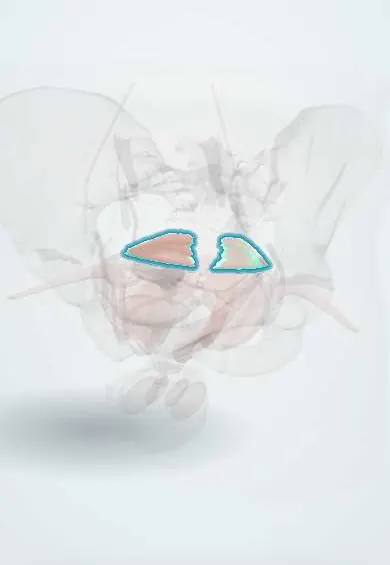
Coccygeus !
The coccygeus is a small muscle at the back of the pelvic floor, between the ischial spine and the tailbone; it completes the floor and steadies the coccyx.
Official name: Coccygeus muscle
Opens in the Durgical app, no sign-up needed.
What it is
A triangular muscle behind the levator ani, from the ischial spine to the sacrum and coccyx, on the sacrospinous ligament.
What it does
Completes the back of the pelvic floor and steadies the coccyx.
Key facts
- Origin
- Ischial spine and sacrospinous ligament
- Insertion
- Lateral margins of the lower sacrum and coccyx
- Action
- Supports the pelvic floor posteriorly; flexes and steadies the coccyx
- Nerve
- Anterior rami of S3–S4
- Blood supply
- Inferior gluteal artery
Relations
- Above
- Piriformis, separated from it by the lower part of the greater sciatic foramen
- In front
- Posterior border of the iliococcygeus, which it continues backwards in the same plane
- Behind
- Sacrospinous ligament on its outer (gluteal) surface; just behind the ligament near the ischial spine cross the pudendal nerve and the internal pudendal vessels
- Medial
- Its pelvic surface faces the rectum and carries the coccygeal plexus
How to find it
It cannot be felt from outside. On vaginal or rectal examination the ischial spine is felt on the side wall, and the firm coccygeus–sacrospinous band can be traced from it back to the side of the sacrum and coccyx. In dissection it is seen from the pelvis just behind the iliococcygeus.
On imaging
Seen on axial and coronal MRI or CT as a thin triangular muscle on the pelvic side of the sacrospinous ligament, from the ischial spine to the edge of the sacrum and coccyx. It is rarely assessed alone; the ischial spine and ligament are the landmarks, and MRI shows the pudendal vessels and nerve passing just behind the spine. Radiographs show only its bony attachments.
Common problems
- Stretching or tearing in vaginal birth.
- Pelvic organ prolapse.
- Urinary or faecal incontinence.
- Overactive, painful pelvic floor (pain with sex, pelvic pain).
How it is diagnosed
Pelvic examination, sometimes ultrasound or MRI of the pelvic floor.
Treatment
- Pelvic floor muscle training (Kegel exercises) with a physiotherapist.
- A pessary (a removable support placed in the vagina).
- Surgery for prolapse that bothers the person.
Clinical notes
- Sacrospinous ligament fixation for uterine or vaginal vault prolapse places sutures in the coccygeus–sacrospinous complex medial to the ischial spine, away from the pudendal nerve and vessels behind the spine
- Tenderness or spasm of the coccygeus and levator ani is one recognised source of coccyx pain (coccydynia) and chronic pelvic pain; it is felt as a tender band beside the coccyx on internal examination
- The pudendal nerve can be trapped where it passes between the sacrospinous ligament (fused with this muscle) and the sacrotuberous ligament, one cause of burning perineal pain that worsens on sitting (pudendal neuralgia)
Anatomical variants
- The amount of muscle varies: in some people it is mostly fibrous and hard to separate from the sacrospinous ligament; MRI can show it thin or absent in some women
- Its nerve may come directly from S4 or from the coccygeal plexus
Development
Its other name, ischiococcygeus, recalls its role in tailed mammals, where it swings the tail to the side; in humans the tail is lost and the muscle is reduced, which is why it is often partly fibrous.
Memory aid
The human «tail-wagger»: in tailed mammals this muscle swings the tail; in humans it is reduced and mainly steadies the coccyx.
Test yourself
Think of the answer, then tap the question to see it.
Is the coccygeus part of the levator ani?
No; with the levator ani it forms the pelvic diaphragm, but it is a separate posterior muscle.
With which ligament is the coccygeus fused, and what is the combined structure called surgically?
The sacrospinous ligament; surgeons treat them as one coccygeus–sacrospinous complex.
In sacrospinous ligament fixation, where are sutures placed relative to the ischial spine, and why?
Medial to the ischial spine, away from the pudendal nerve and internal pudendal vessels that cross behind the spine.
Educational information, not medical advice. For any symptom, see a health professional.
Its names
- Official Latin (TA2)
- musculus coccygeus TA2 2412
- Official Spanish (DeCS)
- Músculo coccígeo DeCS D017773
- English (TA2)
- Coccygeus muscle
- Also called
- ischiococcygeus muscle · musculus coccygicus
Related
Sources: Terminologia Anatomica (TA2); DeCS; Anatomy, Abdomen and Pelvis: Levator Ani Muscle; Anatomy, Abdomen and Pelvis: Pelvic Floor.
Spotted a mistake?We are always improving for the community. Tell us what is wrong and our team will review it.Report a mistake
Representative model, for educational purposes only.