Conditions · Infectious and tropical diseases
Lymphatic filariasis !
A mosquito-borne worm that lives in the lymph vessels and, over years, can cause swollen legs (elephantiasis) and a swollen scrotum (hydrocele).
Updated 2026-11-01
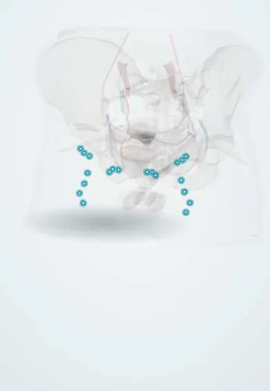
Structures involved
Tap one to see what it is and open it in 3D.
Microorganisms that cause it
Why it happens
- Edema and effusions: the four mechanisms
- Fibrosis in organs: TGF-β and the myofibroblast
- How the lymphatic system works
- Lymphatic vessels
What it is
A neglected tropical disease caused by thread-like filarial worms, mainly Wuchereria bancrofti, that live in the lymphatic vessels. Most infections are silent for years, but they damage lymph drainage and can lead to lymphedema and elephantiasis of the limbs, hydrocele, chyluria and repeated attacks of inflammation.
Who gets it
Hundreds of millions of people in tropical Africa, Asia and the Pacific live in areas that need mass drug treatment, and tens of millions have chronic disease. In the Americas only W. bancrofti occurs, transmitted by Culex mosquitoes; Haiti, the Dominican Republic and Guyana remain endemic, while Brazil was validated as having eliminated it as a public health problem in 2024. In Colombia it is mostly seen as an imported or historical disease, but it is examined as a cause of elephantiasis and hydrocele.
Causes
- Wuchereria bancrofti (about 90% of cases worldwide and the only species in the Americas)
- Brugia malayi and B. timori in South and Southeast Asia
- Transmitted by mosquitoes (Culex, Anopheles, Aedes) that pick up microfilariae from the blood of an infected person
- Repeated bites over months or years are needed, so short-term travellers rarely get it
Risk factors
- Long-term residence in endemic areas with poor sanitation that favours Culex breeding
- Lack of bed nets and mosquito control
- Living in communities that missed mass drug administration rounds
Pathophysiology
Infective larvae deposited by the mosquito migrate to lymphatic vessels and nodes, where they mature into adult worms that live for several years. Adults release microfilariae, which in W. bancrofti usually circulate in the blood at night (nocturnal periodicity). Living adults dilate lymphatic vessels; dying worms and their Wolbachia endosymbiotic bacteria trigger inflammation. Over time lymph flow fails, protein-rich fluid accumulates in the tissues, and repeated secondary bacterial skin infections drive fibrosis, skin thickening and elephantiasis. Obstruction of scrotal lymphatics causes hydrocele, and rupture of lymphatics into the urinary tract causes chyluria.
Classification
- Asymptomatic (subclinical) infection with microfilaremia and hidden lymphatic damage
- Acute: adenolymphangitis attacks (fever, painful nodes, lymphangitis spreading away from the node) and filarial fever
- Chronic: lymphedema graded up to elephantiasis, hydrocele, chyluria
- Tropical pulmonary eosinophilia: immune hyperresponse in the lungs with very high eosinophils
Symptoms and signs
- Often none for years
- Episodes of fever with painful, swollen lymph nodes and red streaks along a limb
- Progressive swelling of a leg, arm, breast or genitals that starts soft and becomes hard and thickened
- Scrotal swelling (hydrocele) in men
- Milky urine (chyluria)
- Nocturnal cough and wheeze with eosinophilia (tropical pulmonary eosinophilia)
Diagnosis
- Clinical
- Suspected in a person from an endemic area with lymphedema, hydrocele or recurrent adenolymphangitis. In late elephantiasis the worms are often dead and parasite tests may be negative, so diagnosis rests on exposure and clinical findings.
- Tests
- Rapid tests for circulating filarial antigen of W. bancrofti, which work at any time of day and are used to map and monitor programmes. Thick blood smear or membrane filtration of blood drawn around midnight to find microfilariae. Eosinophil count and IgE (very high in tropical pulmonary eosinophilia). Testing for onchocerciasis and loiasis exposure before treatment in Africa.
- Imaging
- Scrotal or breast ultrasound can show moving adult worms in dilated lymphatics (the 'filarial dance sign'). Lymphoscintigraphy shows abnormal lymph flow. Chest radiograph shows diffuse nodular infiltrates in tropical pulmonary eosinophilia.
Differential diagnosis
- Non-infectious lymphedema (congenital, after surgery or radiotherapy, malignancy)
- Podoconiosis (non-filarial elephantiasis from irritant volcanic soils)
- Chronic venous insufficiency and heart, kidney or liver causes of edema
- Hydrocele or hernia from other causes
- Recurrent bacterial cellulitis
Goal of treatment
Kill the parasite to stop transmission and further damage, prevent acute attacks and progression of lymphedema through daily limb care, and correct hydrocele surgically.
Treatment
- Medical
- Antifilarial drugs: diethylcarbamazine with albendazole, or ivermectin with albendazole where onchocerciasis coexists (diethylcarbamazine is avoided there because it causes severe reactions); doxycycline over several weeks targets the Wolbachia bacteria and harms adult worms. Mass drug administration of these combinations is the basis of elimination programmes.
- Conservative
- Lymphedema care package: daily washing and drying of the limb, skin and nail care, treatment of fungal infections and wounds, limb elevation, exercise, suitable footwear and compression where tolerated, plus prompt antibiotics for acute attacks.
- Surgical
- Hydrocelectomy for filarial hydrocele; reconstructive lymphatic surgery in selected cases of advanced elephantiasis.
- Follow-up
- Nursing priorities: teaching self-care and skin inspection, watching for cellulitis, measuring limb circumference, and attending to stigma, mental health and disability.
Complications
- Elephantiasis with disability and loss of work
- Recurrent bacterial cellulitis and sepsis
- Hydrocele with sexual and social impact
- Chyluria with protein and fat loss
- Tropical pulmonary eosinophilia leading to lung fibrosis if untreated
- Stigma, depression and social exclusion
Prognosis
Treatment clears microfilariae and can stop early lymphatic damage. Established elephantiasis does not reverse, but consistent hygiene and limb care greatly reduce acute attacks and slow progression. Hydrocele is cured by surgery.
Prevention
- Annual mass drug administration to whole at-risk communities
- Bed nets, repellents and control of Culex breeding sites
- Improved sanitation and drainage
- Morbidity management and disability prevention for people already affected
Test yourself
Think of the answer, then tap the question to see it.
Which filarial worm causes lymphatic filariasis in the Americas?
Wuchereria bancrofti, transmitted mainly by Culex mosquitoes.
Why are blood samples for microfilariae drawn around midnight?
W. bancrofti microfilariae show nocturnal periodicity and circulate in peripheral blood mainly at night.
Which antifilarial drug must be avoided where onchocerciasis coexists?
Diethylcarbamazine, because it causes severe reactions in onchocerciasis (ivermectin plus albendazole is used instead).
What is the 'filarial dance sign'?
Movement of live adult worms seen on ultrasound in dilated scrotal or breast lymphatics.
What is the main measure that prevents acute attacks in lymphedema?
Daily limb hygiene and skin care with prompt treatment of skin infections.
Related conditions
Educational information, not medical advice. For any symptom, see a health professional.
Sources: Lymphatic filariasis (World Health Organization, 2025); Filariasis linfática - OPS/OMS | Organización Panamericana de la Salud (Organización Panamericana de la Salud, 2025).
Spotted a mistake?We are always improving for the community. Tell us what is wrong and our team will review it.Report a mistake
Representative model, for educational purposes only.