Muscular system · Head and neck
Face fasciae !
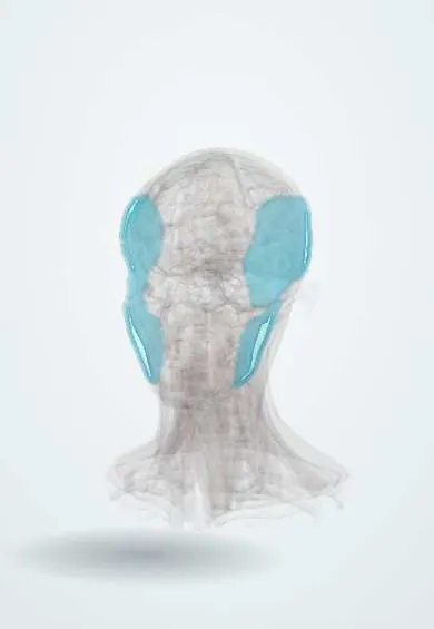
The temporal and masseteric fasciae are thin sheets covering the chewing muscles of the temple and jaw; the parotid gland rests on them.
Official name: Superficial layer of temporal fascia
Opens in the Durgical app, no sign-up needed.
What it is
Tough sheets over the temporalis and masseter.
What it does
Cover the chewing muscles; the parotid sits on the masseteric fascia.
Key facts
- Attaches
- Temporal fascia: superior temporal line to the zygomatic arch; masseteric fascia: zygomatic arch to the mandible, continuous with the parotid fascia
- Function
- Cover the temporalis and masseter
Relations
- Superficial
- Subcutaneous fat and skin, tied to the deeper layers by retaining ligaments (zygomatic and mandibular ligaments)
- Deep
- Masseter and temporalis, the parotid gland and duct, the buccal fat pad, and the facial nerve branches
- Above
- Temporal fascia attached to the superior temporal line; the temporoparietal fascia and galea over it
- Below
- Platysma and the investing layer of the cervical fascia over the mandible
How to find it
Not palpable as a layer. In a facelift or parotid approach the SMAS is the first firm layer under the subcutaneous fat; the temporal fascia is the shiny white sheet felt on the temple under the scalp layers.
On imaging
Ultrasound and MRI show the SMAS as a thin line between the subcutaneous fat and the parotid or masseter; the temporal fat pad lies between the layers of the temporal fascia above the zygomatic arch.
Clinical notes
- Facelift techniques tighten, fold or lift the SMAS rather than the skin alone; the deeper the plane, the closer the dissection comes to the facial nerve branches
- Gillies approach for a depressed zygomatic arch fracture: an incision in the temporal hairline, then an elevator passed deep to the temporal fascia, on the temporalis, under the arch to lift it
- In the coronal approach the superficial layer of the deep temporal fascia is opened above the arch, keeping the temporal branch in the flap
- Mumps and parotid infection are painful because the gland swells inside its unyielding fascia
Test yourself
Think of the answer, then tap the question to see it.
What is the SMAS, and which nerve branches lie deep to it?
The superficial musculoaponeurotic system, one fibromuscular layer joining the platysma to the temporoparietal fascia and galea; the facial nerve branches lie deep to it.
Why does a swollen parotid gland, as in mumps, hurt?
The masseteric fascia continues as the parotid fascia, a tight capsule, so the gland swells inside an unyielding sheath.
In the Gillies approach for a depressed zygomatic arch fracture, in which plane is the elevator passed?
Deep to the temporal fascia, on the temporalis, from an incision in the temporal hairline, then under the arch to lift it.
Educational information, not medical advice. For any symptom, see a health professional.
Its names
- Official Latin (TA2)
- lamina superficialis fasciae temporalis TA2 2139
- Official Spanish (DeCS)
- Fascias temporal y maseterina
- English (TA2)
- Superficial layer of temporal fascia
Sources: Terminologia Anatomica (TA2); Anatomy, Skin, Superficial Musculoaponeurotic System (SMAS) Fascia; Anatomy, Head and Neck: Face.
Spotted a mistake?We are always improving for the community. Tell us what is wrong and our team will review it.Report a mistake
Representative model, for educational purposes only.