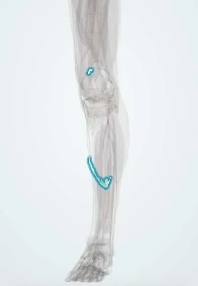
Gastrocnemius tendon (inner head) !
The inner gastrocnemius tendon anchors the inner head of the calf muscle to the back of the femur, just above the knee.
Official name: Tendon of medial head of gastrocnemius
Opens in the Durgical app, no sign-up needed.
What it is
The upper tendon of the inner calf muscle head, on the back of the femur.
What it does
Anchors the calf muscle above the knee.
Key facts
- Attaches
- Origin tendon of the medial head on the popliteal surface of the femur, just above the medial condyle and onto the posterior capsule
- Function
- Anchors the medial head above the knee, so the gastrocnemius flexes the knee as well as plantarflexing the ankle; forms the inferomedial border of the popliteal fossa
- Tight in
- Knee extension with ankle dorsiflexion
Relations
- Lateral
- Popliteal artery, popliteal vein and tibial nerve, which pass down between the two heads
- Medial
- Semimembranosus tendon, with the semimembranosus–gastrocnemius bursa between them
- Deep
- Posterior knee joint capsule over the medial femoral condyle, with a small bursa beneath the tendon
- Superficial
- Popliteal fascia and skin; the small saphenous vein ends in the fossa between the two heads
How to find it
Prone with the knee slightly bent and the ball of the foot pressed down, the medial head forms the inner lower border of the popliteal fossa; its upper tendon lies deep above the medial condyle and is hard to feel.
Examination
- Foucher sign: a swelling at the back of the knee that is tense with the knee straight and softens when it is bent is typical of a popliteal (Baker's) cyst; a mass that stays firm in both positions needs further imaging
On imaging
On axial ultrasound or MRI at the back of the knee a Baker's cyst shows a neck between this tendon and the semimembranosus; in suspected artery entrapment, imaging with the ankle in active plantarflexion shows the artery's course relative to the medial head.
Clinical notes
- In popliteal artery entrapment, surgery releases the abnormal muscle slip or tendon and repairs the artery if it is damaged
- The medial gastrocnemius flap pivots on this upper tendon, where its pedicle (the medial sural artery) enters; detaching the origin from the femur lengthens the arc of rotation
- Arthroscopic treatment of a popliteal cyst works at the posteromedial capsule, beside this tendon, where the valve-like opening of the cyst lies; the knee problem that feeds the cyst is treated at the same time
Anatomical variants
- A third head of gastrocnemius (gastrocnemius tertius) can arise from the popliteal surface of the femur and may entrap the popliteal artery
Related conditions
Test yourself
Think of the answer, then tap the question to see it.
Between which two tendons is the neck of a Baker's cyst found?
Between the medial head of gastrocnemius tendon and the semimembranosus tendon.
What is popliteal artery entrapment?
Compression of the popliteal artery by an abnormal course medial to the medial head of gastrocnemius, or an abnormal slip, causing exercise calf pain in young athletes.
Where is the tear in 'tennis leg'?
At the lower end of the medial head of gastrocnemius, not at its upper tendon.
Educational information, not medical advice. For any symptom, see a health professional.
Its names
- Official Latin (TA2)
- caput mediale musculi gastrocnemii TA2 2659
- Official Spanish (DeCS)
- Tendón del gemelo interno DeCS D013710
- English (TA2)
- Tendon of medial head of gastrocnemius
- Also called
- caput tibiale musculi gastrocnemii
Related
Sources: Terminologia Anatomica (TA2); DeCS; Anatomy, Bony Pelvis and Lower Limb: Gastrocnemius Muscle.
Spotted a mistake?We are always improving for the community. Tell us what is wrong and our team will review it.Report a mistake
Representative model, for educational purposes only.