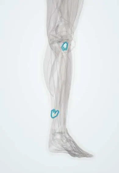
Soleus tendon !
The soleus tendon is the tendon part of the deep calf muscle; it joins the gastrocnemius to form the Achilles tendon.
Official name: Tendon of soleus
Opens in the Durgical app, no sign-up needed.
What it is
The tendon part of the soleus, the deep calf muscle.
What it does
Joins the Achilles tendon to point the foot.
Key facts
- Attaches
- Aponeurosis on the back of the soleus that fuses with the gastrocnemius aponeurosis to form the calcaneal (Achilles) tendon, on the middle of the posterior calcaneus
- Function
- Plantarflexion whatever the knee position (main plantarflexor in standing and slow walking); its contraction drives the calf muscle pump
- Tight in
- Ankle dorsiflexion with the knee flexed
Relations
- Superficial
- Gastrocnemius aponeurosis, with the plantaris tendon between them
- Deep
- Deep transverse fascia, then the deep compartment: tibialis posterior, flexor digitorum longus, flexor hallucis longus, posterior tibial artery and tibial nerve
- Below
- Merges into the Achilles tendon; Kager's fat pad lies in front of the tendon above the calcaneus
How to find it
Rising onto tiptoe with the knees bent brings out the soleus on each side below the gastrocnemius; its flat aponeurosis is felt where the calf narrows into the Achilles tendon.
Examination
- Silfverskiöld test: ankle dorsiflexion is measured with the knee straight and bent; if it improves with the knee bent, the tightness is in the gastrocnemius; if it stays limited, the soleus and Achilles tendon are tight
On imaging
On ultrasound and MRI its aponeurosis is a dark band deep to the gastrocnemius; soleus strains are often centred on the central or side aponeuroses and are easier to see on MRI.
Clinical notes
- Surgical lengthening for a tight calf is chosen by level: gastrocnemius recession spares the soleus, while Achilles tendon lengthening affects both
- Tears where the soleus aponeurosis joins the Achilles tendon (a musculotendinous injury) are usually treated without surgery, unlike some ruptures within the tendon itself
Related conditions
Test yourself
Think of the answer, then tap the question to see it.
How does the Silfverskiöld test tell soleus tightness from gastrocnemius tightness?
If dorsiflexion improves with the knee bent, the gastrocnemius is tight; if it stays limited, the soleus and Achilles are tight.
What does the soleus aponeurosis form, and where does it insert?
It fuses with the gastrocnemius aponeurosis to form the calcaneal (Achilles) tendon, on the middle of the posterior calcaneus.
What passes under the fibrous arch of the soleus?
The popliteal artery and the tibial nerve, entering the deep calf.
Educational information, not medical advice. For any symptom, see a health professional.
Its names
- Official Latin (TA2)
- musculus soleus TA2 2660
- Official Spanish (DeCS)
- Tendón del sóleo DeCS D013710
- English (TA2)
- Tendon of soleus
Related
Sources: Terminologia Anatomica (TA2); DeCS; Anatomy, Bony Pelvis and Lower Limb: Calf; Anatomy, Bony Pelvis and Lower Limb: Leg Posterior Compartment.
Spotted a mistake?We are always improving for the community. Tell us what is wrong and our team will review it.Report a mistake
Representative model, for educational purposes only.