Cardiovascular system · Lower limb
Posterior tibial artery !
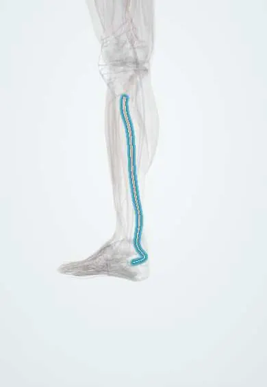
The posterior tibial artery runs down the back of the leg and behind the inner ankle to the sole; its pulse is felt there.
Opens in the Durgical app, no sign-up needed.
What it is
The main artery of the back of the leg, passing behind the inner ankle bone to the sole.
What it does
Supplies the calf muscles and, through its plantar branches, the sole of the foot.
Key facts
- Origin
- Popliteal artery at lower border of popliteus; its first segment, before the fibular artery arises, is the tibioperoneal (tibiofibular) trunk
- Branches
- Fibular artery, nutrient artery of tibia, medial malleolar, calcaneal; ends as medial and lateral plantar arteries
- Supplies
- Posterior compartment of leg and sole of foot; pulse behind medial malleolus
Relations
- Course
- From the lower border of the popliteus it passes under the soleus arch, runs down the deep posterior compartment on the tibialis posterior and then the flexor digitorum longus, reaches the space between the medial malleolus and the heel, and divides under the abductor hallucis into the plantar arteries
- Superficial
- The gastrocnemius and soleus, separated from it by the deep transverse fascia; above the ankle only skin, fascia and the flexor retinaculum
- Deep
- The tibialis posterior and flexor digitorum longus, then the back of the tibia and the ankle joint
- Lateral
- The tibial nerve: medial to the artery at the top, it crosses behind it and lies lateral to it for most of the leg
How to find it
With the person supine and the foot relaxed, the fingertips curl behind the medial malleolus, halfway toward the heel, and press lightly into the soft hollow there.
Examination
- Ankle–brachial index: the higher ankle pressure (posterior tibial or dorsalis pedis) divided by the higher arm pressure; 0.90 or less indicates peripheral artery disease, above about 1.40 suggests stiff, calcified arteries
- Handheld Doppler: a normal signal is triphasic; a monophasic signal points to narrowing upstream
- Buerger test: the foot turns pale when the leg is raised and dusky red when it hangs down, a sign of severe ischaemia
On imaging
Duplex ultrasound maps narrowings from the calf down to the ankle. CT or catheter angiography shows whether it stays open into the plantar arteries before a bypass. In diabetes or kidney disease calcified walls can blur the images and falsely raise its pressure.
Common problems
- Its pulse behind the inner ankle is checked to assess blood flow to the foot.
- Narrowing from peripheral artery disease can cause calf pain on walking.
Clinical notes
- Tarsal tunnel syndrome affects the tibial nerve beside it; enlarged companion veins are one cause
- A frequent target for distal bypass in limb-threatening ischemia
- Medial malleolus fractures, their screws and medial ankle surgery lie close to it
- After a long ischaemia, swelling of the deep posterior compartment around it when flow returns can cause compartment syndrome
- In the angiosome model it feeds the heel and most of the sole, so healing of a heel ulcer depends largely on it
Anatomical variants
- Hypoplastic or absent, with the fibular artery continuing to the heel and sole
- High origin when the popliteal artery divides above the usual level
Development
It buds from the embryonic axis artery of the leg (the sciatic artery and its continuation, the fibular artery). This shared origin is why the fibular artery can take over the heel and sole when the posterior tibial artery is small.
Memory aid
Tarsal tunnel, front to back: «Tom, Dick And Very Nervous Harry»: Tibialis posterior, flexor Digitorum longus, Artery, Vein, Nerve (tibial), flexor Hallucis longus.
Related conditions
Test yourself
Think of the answer, then tap the question to see it.
Where is the posterior tibial pulse felt?
Behind the medial malleolus, halfway between it and the calcaneal tuberosity, under the flexor retinaculum.
Which is more significant clinically: an absent posterior tibial or an absent dorsalis pedis pulse?
An absent posterior tibial pulse; a missing dorsalis pedis pulse can be a normal variant.
How is the ankle–brachial index calculated, and which values are abnormal?
Higher ankle pressure (posterior tibial or dorsalis pedis) over higher arm pressure; 0.90 or less means artery disease, above about 1.40 stiff arteries.
What are the main branches and terminal branches of the posterior tibial artery?
Fibular artery, tibial nutrient, medial malleolar and calcaneal branches; it ends as the medial and lateral plantar arteries.
What does the Buerger test show in severe leg ischaemia?
The foot goes pale when the leg is raised and dusky red when it hangs down.
Educational information, not medical advice. For any symptom, see a health professional.
Its names
- Official Latin (TA2)
- arteria tibialis posterior TA2 4721
- Official Spanish (DeCS)
- Arteria tibial posterior DeCS D016909
- English (TA2)
- Posterior tibial artery
Related
Sources: Terminologia Anatomica (TA2); DeCS; Anatomy, Bony Pelvis and Lower Limb: Posterior Tibial Artery; 20.5 Circulatory Pathways (OpenStax (Rice University), 2022) · adapted, CC BY 4.0; Anatomy of the Human Body (20th ed.): Development of the Vascular System (Lea & Febiger (Gray's Anatomy, 1918), 1918) · adapted, Public domain.
Spotted a mistake?We are always improving for the community. Tell us what is wrong and our team will review it.Report a mistake
Representative model, for educational purposes only.