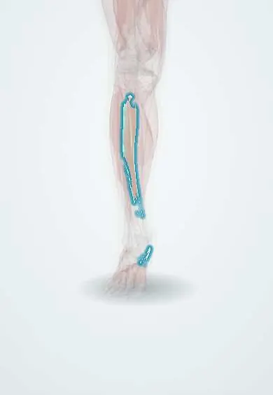
Tibialis posterior !
The tibialis posterior is a deep calf muscle whose tendon runs behind the inner ankle; it supports the arch of the foot.
Official name: Tibialis posterior muscle
Opens in the Durgical app, no sign-up needed.
What it is
A deep calf muscle lying behind the tibia, the fibula and the membrane between them. Its tendon passes behind the inner ankle to the arch.
What it does
Supports the arch of the foot, turns the foot inward and helps point it.
Key facts
- Origin
- Interosseous membrane; posterior tibia below soleal line; posterior fibula
- Insertion
- Navicular tuberosity; cuneiforms; cuboid; bases of metatarsals 2–4
- Action
- Plantar flexes ankle; inverts foot; supports medial longitudinal arch
- Nerve
- Tibial nerve (L4–L5)
- Blood supply
- Posterior tibial artery; fibular artery
Relations
- Course
- Its tendon grooves the back of the medial malleolus, runs under the flexor retinaculum, passes below the malleolus over the spring ligament and fans out to the navicular and the other tarsal and metatarsal bones
- In front
- Interosseous membrane and the back of the tibia and fibula
- Behind
- Deep transverse fascia and soleus; the posterior tibial vessels and tibial nerve run down on its surface
- Medial
- Flexor digitorum longus, which crosses superficial to it just above the ankle
- Lateral
- Flexor hallucis longus, with the fibular vessels near the fibula
- Below
- Spring (plantar calcaneonavicular) ligament, which it supports under the head of the talus
How to find it
With the foot plantarflexed and turned in against resistance, its tendon stands out just behind and below the medial malleolus and can be followed to the navicular tuberosity.
Examination
- Single-leg heel raise: normally the heel swings into varus as it lifts; inability to rise, or a heel that stays in valgus, indicates tibialis posterior dysfunction
- "Too many toes" sign: viewed from behind with the person standing, more lateral toes are visible on the affected side because the forefoot is abducted and the heel everted
- Resisted inversion with the foot plantarflexed: pain or weakness along the tendon
On imaging
Ultrasound and MRI show its tendon as the first and largest behind the medial malleolus, roughly twice the size of the flexor digitorum longus. In dysfunction, look for thickening, fluid, split tears or rupture behind and just below the malleolus, and an accessory navicular at its insertion. Weight-bearing radiographs show the flatfoot: a sagging talus–first metatarsal line on the lateral view.
Common problems
- Tendon dysfunction: the tendon becomes inflamed, stretched or torn, usually in middle-aged adults. It is the most common cause of adult-acquired flatfoot.
- Pain and swelling along the inner ankle, and trouble standing on tiptoe.
- Rarely, a sudden tear or the tendon slipping out of its groove.
How it is diagnosed
A physical exam, including a single heel rise; standing X-rays; MRI or ultrasound of the tendon.
Treatment
- Rest, a boot or brace, arch supports and physical therapy.
- Surgery to clean or repair the tendon, often with a transfer of a nearby toe-flexor tendon and bone cuts to realign the foot.
- Advanced, stiff flatfoot: fusion of the joints of the back of the foot.
Clinical notes
- Deep posterior compartment syndrome (tibialis posterior, flexor digitorum longus, flexor hallucis longus, posterior tibial and fibular vessels, tibial nerve): pain on passive toe extension and numbness of the sole; easily missed because the compartment is deep
- An accessory navicular can take part of its insertion and cause medial midfoot pain
- Medial tibial stress syndrome (shin splints) along the posteromedial tibial border is linked to traction by tibialis posterior, soleus or flexor digitorum longus; texts differ on which
- In flexible flatfoot from its failure, the flexor digitorum longus tendon can be transferred to the navicular to replace it
Anatomical variants
- Its insertion may be three bands (main, plantar and recurrent) or a single broad tendon
- An accessory navicular may be present at its insertion
Development
From the ventral (flexor) muscle mass of the deep calf, supplied by the tibial nerve; an accessory navicular, from an ossification centre at the navicular tuberosity that fails to fuse, may take part of its insertion.
Memory aid
Behind the medial malleolus, front to back: "Tom, Dick And Very Nervous Harry" (Tibialis posterior, flexor Digitorum longus, posterior tibial Artery, Vein, tibial Nerve, flexor Hallucis longus).
Related conditions
Test yourself
Think of the answer, then tap the question to see it.
What is the order of structures behind the medial malleolus?
Tibialis posterior, flexor digitorum longus, posterior tibial artery, vein, tibial nerve, flexor hallucis longus («Tom, Dick And Very Nervous Harry»).
What are the actions and nerve supply of the tibialis posterior?
It plantar flexes the ankle, inverts the foot and supports the medial longitudinal arch; it is supplied by the tibial nerve (L4–L5).
Which condition is the most common cause of adult-acquired flatfoot?
Tibialis posterior tendon dysfunction, usually starting in the poorly supplied zone behind and just below the medial malleolus.
What does the «too many toes» sign show?
Seen from behind, more lateral toes are visible on the affected side because the forefoot is abducted and the heel everted: tibialis posterior dysfunction.
Which single-leg heel raise finding points to tibialis posterior dysfunction?
Inability to rise, or a heel that stays in valgus instead of swinging into varus as it lifts.
Educational information, not medical advice. For any symptom, see a health professional.
Its names
- Official Latin (TA2)
- musculus tibialis posterior TA2 2666
- Official Spanish (DeCS)
- Músculo tibial posterior
- English (TA2)
- Tibialis posterior muscle
Related
Sources: Terminologia Anatomica (TA2); Anatomy, Bony Pelvis and Lower Limb: Tibialis Posterior Muscle.
Spotted a mistake?We are always improving for the community. Tell us what is wrong and our team will review it.Report a mistake
Representative model, for educational purposes only.