Conditions · Gastroenterology and hepatology
Barrett oesophagus !
A change in the lining of the lower oesophagus caused by long-standing acid reflux. It raises the risk of oesophageal adenocarcinoma.
Updated 2026-11-01
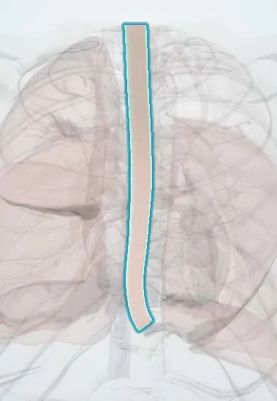
Structures involved
Tap one to see what it is and open it in 3D.
Why it happens
- Metaplasia
- Precancerous conditions: when chronic inflammation, metaplasia or hyperplasia raise the risk
- Dysplasia and carcinoma in situ
- Multistep carcinogenesis: the adenoma–carcinoma sequence
- Biopsy: excisional, incisional, core needle, punch and fine-needle aspiration
- Special stains in diagnosis: Congo red, Prussian blue, PAS, Ziehl–Neelsen, Gram
Show 4 more
What it is
Replacement of the normal stratified squamous lining of the distal oesophagus by columnar epithelium with intestinal metaplasia (goblet cells), as a result of chronic gastro-oesophageal reflux. It is the main precursor of oesophageal adenocarcinoma.
Who gets it
Found in a minority of people with chronic reflux, most often white middle-aged and older men with central obesity. In Colombia and Latin America, gastric cancer remains far more common than oesophageal adenocarcinoma, but reflux and obesity are rising, so Barrett oesophagus is increasingly found at endoscopy.
Causes
- Chronic gastro-oesophageal reflux disease (acid and bile exposure of the distal oesophagus)
- Metaplasia: the injured squamous lining heals with a columnar, acid-resistant intestinal-type epithelium
Risk factors
- Reflux symptoms for five years or more
- Male sex and age over 50
- Central (abdominal) obesity
- Smoking
- Hiatal hernia
- Family history of Barrett oesophagus or oesophageal adenocarcinoma
Pathophysiology
Repeated acid and bile injury damages the squamous mucosa of the distal oesophagus. Healing takes place with columnar cells containing goblet cells (intestinal metaplasia), which resist acid better but are genetically unstable. The metaplasia can progress through low-grade and high-grade dysplasia to invasive adenocarcinoma, usually over many years.
Classification
- By length: short-segment (under 3 cm) or long-segment (3 cm or more), described with the Prague C and M criteria
- By histology: non-dysplastic, indefinite for dysplasia, low-grade dysplasia, high-grade dysplasia, intramucosal carcinoma
Symptoms and signs
- No symptoms of its own; it is found during endoscopy for reflux
- Heartburn and acid regurgitation from the underlying reflux
- Dysphagia, weight loss or bleeding suggest stricture or cancer
Diagnosis
- Clinical
- Suspected in people with chronic reflux, especially men over 50 with additional risk factors; screening endoscopy is considered in this group.
- Tests
- Upper endoscopy shows salmon-coloured mucosa extending above the gastro-oesophageal junction. Diagnosis requires biopsies showing intestinal metaplasia with goblet cells; systematic four-quadrant biopsies look for dysplasia, which an expert pathologist confirms.
- Imaging
- Not needed for diagnosis; CT or endoscopic ultrasound stage a cancer when one is found.
Differential diagnosis
- Erosive oesophagitis without metaplasia
- Gastric heterotopia (inlet patch)
- Oesophageal adenocarcinoma
- Eosinophilic oesophagitis
Goal of treatment
Control reflux, find dysplasia early with surveillance endoscopy, and remove dysplastic or early neoplastic tissue before it becomes invasive cancer.
Treatment
- Medical
- Long-term proton pump inhibitor therapy to control reflux and oesophagitis.
- Lifestyle
- Weight loss, stopping smoking, raising the head of the bed and avoiding late meals.
- Follow-up
- Surveillance endoscopy with biopsies at intervals set by length and grade of dysplasia; non-dysplastic Barrett needs the longest intervals.
- Procedure
- Endoscopic eradication for confirmed dysplasia or early cancer: endoscopic resection of visible lesions followed by radiofrequency ablation of the remaining Barrett mucosa.
- Surgical
- Antireflux surgery (fundoplication) for refractory reflux; oesophagectomy is reserved for invasive cancer not suitable for endoscopic treatment.
Complications
- Oesophageal adenocarcinoma
- Peptic stricture
- Barrett ulcer and bleeding
Prognosis
Most people with non-dysplastic Barrett oesophagus never develop cancer and have a normal life expectancy; the yearly risk of progression is low but rises markedly with high-grade dysplasia. Endoscopic eradication is highly effective when dysplasia is found early.
Prevention
- Effective control of chronic reflux
- Healthy weight and no smoking
- Screening endoscopy in high-risk people with chronic reflux
Test yourself
Think of the answer, then tap the question to see it.
What histological change defines Barrett oesophagus?
Intestinal metaplasia: squamous epithelium replaced by columnar epithelium with goblet cells in the distal oesophagus.
Which cancer does Barrett oesophagus predispose to?
Oesophageal adenocarcinoma of the distal oesophagus (not squamous cell carcinoma).
What is the typical patient profile?
A middle-aged or older white man with long-standing reflux, central obesity and a history of smoking.
How is high-grade dysplasia in Barrett oesophagus usually treated?
Endoscopic eradication: resection of visible lesions plus radiofrequency ablation.
Related conditions
- Gastro-oesophageal reflux disease (GERD)
- Hiatal hernia
- Oesophageal cancer
- Eosinophilic oesophagitis
- Peptic ulcer disease
Educational information, not medical advice. For any symptom, see a health professional.
Sources: Barrett esophagus: MedlinePlus Medical Encyclopedia (MedlinePlus (U.S. National Library of Medicine), 2024); Barrett’s Esophagus - NIDDK (National Institute of Diabetes and Digestive and Kidney Diseases (NIDDK), 2024).
Spotted a mistake?We are always improving for the community. Tell us what is wrong and our team will review it.Report a mistake
Representative model, for educational purposes only.