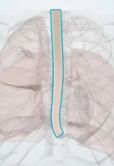
Esophagus !
The esophagus is the muscular tube that carries food from the throat to the stomach, passing down the back of the chest behind the heart.
Opens in the Durgical app, no sign-up needed.
What it is
The muscular food pipe running from the throat, behind the trachea and heart, through the diaphragm to the stomach.
What it does
Moves food down with waves of muscle (peristalsis); a muscular valve at the bottom keeps stomach acid out.
Key facts
- Where
- From C6 (cricoid) behind the trachea, through the diaphragm at T10, to the cardia at about T11; about 25 cm
- Blood supply
- Cervical: inferior thyroid arteries; thoracic: esophageal branches of aorta and bronchial arteries; abdominal: left gastric and left inferior phrenic arteries
- Nerve
- Vagus (recurrent laryngeal nerves above, esophageal plexus below); sympathetic trunk
- Function
- Moves food to the stomach by peristalsis; upper and lower esophageal sphincters; upper third striated, lower third smooth muscle
Relations
- Course
- From the cricoid it descends in the midline behind the trachea, drifts slightly left at the root of the neck, is pushed back to the midline by the aortic arch at T4, then swings forward and left in front of the descending aorta to pass through the diaphragm
- In front
- Trachea and left recurrent laryngeal nerve above; the tracheal bifurcation, left main bronchus and subcarinal nodes; then the pericardium over the left atrium; lowest, the diaphragm
- Behind
- Vertebral column and longus colli, thoracic duct, azygos vein, right posterior intercostal arteries and hemiazygos veins; in the lower chest the descending aorta
- Lateral
- Left: the aortic arch, left subclavian artery, upper thoracic duct and left pleura. Right: the arch of the azygos vein, the right pleura and lung
How to find it
On endoscopy, distances are measured from the incisor teeth: the upper sphincter at about 15 cm and the gastroesophageal junction at about 40 cm, at the Z-line where pale squamous lining meets salmon-pink gastric mucosa.
On imaging
Barium swallow shows the narrowings, the impressions of the aortic arch and left main bronchus, a large left atrium, webs, strictures and the bird's-beak taper of achalasia. CT shows wall thickening, nodes and, after a perforation, mediastinal air and pleural fluid.
Common problems
- GERD (reflux): acid comes back up, causing heartburn.
- Barrett's esophagus: long-term reflux changes the lining, raising cancer risk.
- Hiatal hernia: part of the stomach slides up through the diaphragm.
How it is diagnosed
Symptoms; upper endoscopy; pH monitoring; barium swallow.
Treatment
- Lifestyle changes and acid-reducing medicines; surgery (fundoplication) for some.
Clinical notes
- Esophageal varices at the lower end, where left gastric (portal) and azygos (systemic) veins meet; they can bleed heavily in portal hypertension
- Boerhaave syndrome: full-thickness rupture after forceful vomiting, usually in the left lower posterolateral wall; mediastinal air, left pleural effusion, crunching sound with the heartbeat (Hamman sign)
- Squamous cell carcinoma arises mostly in the upper and middle thirds; adenocarcinoma in the lower third, on Barrett's esophagus
- Esophagectomy (transhiatal or through the right chest) risks chylothorax, recurrent laryngeal nerve injury and anastomotic leak
- Swallowed coins and food boluses stick at the narrowings, most often at the upper sphincter in children
Development
The tracheoesophageal septum splits the foregut into trachea and esophagus; faulty separation causes esophageal atresia and tracheoesophageal fistula, most often an upper blind pouch with a lower fistula (polyhydramnios, frothing and choking at feeds).
Related conditions
- Upper gastrointestinal bleeding
- HIV infection and AIDS
- Chagas disease
- Paraquat poisoning
- Caustic ingestion
- Barrett oesophagus
- Eosinophilic oesophagitis
- Mallory-Weiss syndrome
- Oesophageal perforation (Boerhaave syndrome)
- Hiatal hernia
- Infectious and pill-induced oesophagitis (Candida, HSV, CMV)
- Zenker diverticulum (pharyngo-oesophageal diverticulum)
- Distal oesophageal spasm and hypercontractile oesophagus
- Chagasic megaoesophagus and megacolon
- Cytomegalovirus infection
- Candidiasis: oral, oesophageal and invasive
- Esophageal atresia and tracheoesophageal fistula
- VACTERL association
- Mediastinitis (acute and fibrosing)
- Systemic sclerosis (scleroderma, including CREST)
- Mixed connective tissue disease
- Gastro-oesophageal reflux disease (GERD)
- Achalasia
- Oesophageal cancer
Test yourself
Think of the answer, then tap the question to see it.
What are the three narrowings of the esophagus where swallowed objects lodge?
The cricopharyngeus (about 15 cm from the incisors), the crossing of the aortic arch and left main bronchus (about 22–27 cm) and the diaphragmatic hiatus (about 40 cm).
Which heart chamber lies directly in front of the thoracic esophagus, and why does it matter?
The left atrium: a large left atrium indents the esophagus on a barium swallow, and transesophageal echo views the heart through it.
Why do esophageal varices form at the lower end of the esophagus?
Left gastric (portal) and azygos (systemic) veins meet there; in portal hypertension these veins dilate and can bleed heavily.
What is Boerhaave syndrome, and where does the esophagus usually tear?
Full-thickness rupture after forceful vomiting, usually in the left lower posterolateral wall, with mediastinal air and a left pleural effusion.
Why does esophageal cancer spread outward early and why do esophageal anastomoses leak more easily?
The thoracic esophagus has no serosa, only adventitia.
Educational information, not medical advice. For any symptom, see a health professional.
Its names
- Official Latin (TA2)
- oesophagus; pars thoracica oesophagi; pars abdominalis oesophagi TA2 2887
- Official Spanish (DeCS)
- Esófago DeCS D004947
- English (TA2)
- Esophagus
Related
Sources: Terminologia Anatomica (TA2); DeCS; Anatomy, Thorax, Esophagus.
Spotted a mistake?We are always improving for the community. Tell us what is wrong and our team will review it.Report a mistake
Representative model, for educational purposes only.