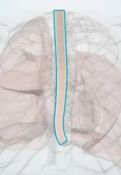
Oesophageal cancer !
Esophageal cancer is a malignant tumour of the esophagus, the food tube; it is often found late, when swallowing becomes difficult and weight drops.
Updated 2026-10-29
Structures involved
Tap one to see what it is and open it in 3D.
- Esophagus
- Mediastinal lymph nodes
- Trachea
- Recurrent laryngeal nerves
- Thoracic aorta
- Main bronchi
- Diaphragm
In biochemistry
In histology
- Cell adaptations: hypertrophy, hyperplasia, atrophy, metaplasia
- The GI tract wall: mucosa, submucosa, muscularis, serosa; enteric plexuses
- Esophagus and the Barrett change
In physiology
Why it happens
- Metaplasia
- Dysplasia and carcinoma in situ
- Diet, cancer and systemic disease
- Multistep carcinogenesis: the adenoma–carcinoma sequence
- Tobacco: how smoking injures tissue
- Chemical carcinogens: initiation, promotion and the table to know
Show 2 more
What it is
A malignant tumour of the oesophagus, mostly squamous cell carcinoma (linked to tobacco and alcohol) or adenocarcinoma (distal, arising from Barrett's oesophagus after chronic reflux). It usually presents late with progressive dysphagia and weight loss.
Who gets it
Squamous cell carcinoma predominates worldwide and in much of Latin America; adenocarcinoma has risen in countries with more obesity and reflux. It is more common in men and older adults and is often diagnosed at an advanced stage.
Causes
- Squamous cell carcinoma: repeated mucosal injury from tobacco, alcohol, very hot drinks and some dietary carcinogens
- Adenocarcinoma: chronic reflux leading to Barrett's oesophagus, then dysplasia and cancer
- Long-standing achalasia or caustic injury (squamous type)
Risk factors
- Smoking
- Heavy alcohol use
- Very hot drinks and a diet low in fruit and vegetables
- Barrett's oesophagus and long-standing reflux disease
- Obesity (adenocarcinoma)
- Male sex and older age
- Achalasia or a previous caustic stricture
Pathophysiology
Repeated injury leads to dysplasia and invasive cancer. The oesophagus has no serosa and a rich lymphatic network, so tumours spread early to nearby and mediastinal nodes and invade the trachea, bronchi, aorta or recurrent laryngeal nerve. Because the oesophagus stretches, dysphagia appears only when the lumen is markedly narrowed.
Classification
- Histology: squamous cell carcinoma or adenocarcinoma
- Site: upper, middle or lower thoracic oesophagus, or gastro-oesophageal junction
- TNM stage
Symptoms and signs
- Progressive dysphagia, first to solids and then to liquids
- Unintended weight loss
- Painful swallowing and pain behind the sternum
- Regurgitation
- Hoarseness from recurrent laryngeal nerve involvement
- Cough when swallowing (tracheo-oesophageal fistula)
- Iron-deficiency anaemia or bleeding
Diagnosis
- Clinical
- Progressive dysphagia with weight loss, especially in a smoker or drinker or in someone with long-standing reflux.
- Tests
- Upper endoscopy with biopsy confirms the diagnosis and the type. Blood tests look for anaemia and assess nutrition. Bronchoscopy is used for upper and middle tumours near the airway.
- Imaging
- CT of the chest and abdomen and PET-CT stage the disease; endoscopic ultrasound assesses depth and nodes. A barium swallow may show an irregular narrowing.
Differential diagnosis
- Benign peptic stricture
- Achalasia
- Eosinophilic oesophagitis
- Cancer of the gastric cardia
- Extrinsic compression by a mediastinal mass
- Oesophageal ring or web
Goal of treatment
Cure localised disease with combined treatment, and in advanced disease relieve dysphagia, maintain nutrition and control symptoms.
Treatment
- Procedure
- Endoscopic resection for very early, superficial tumours, and endoscopic treatment of dysplastic Barrett's oesophagus.
- Surgical
- Oesophagectomy, often after chemoradiotherapy or chemotherapy, for resectable locally advanced disease.
- Medical
- Chemotherapy, definitive chemoradiotherapy (especially for squamous tumours) and, in selected cases, targeted therapy or immune checkpoint inhibitors.
- Procedure
- Palliation: a self-expanding stent to relieve dysphagia and feeding tubes for nutritional support.
- Follow-up
- Endoscopic surveillance of Barrett's oesophagus to find dysplasia early.
- Lifestyle
- Stopping smoking and alcohol, with nutritional support throughout treatment.
Complications
- Malnutrition and cachexia
- Aspiration pneumonia
- Tracheo-oesophageal fistula
- Bleeding
- Anastomotic leak or stricture after oesophagectomy
- Spread to the liver, lungs and bones
Prognosis
Often poor, because most cancers are found late. Early tumours treated by endoscopy or surgery have much better survival.
Prevention
- Avoiding tobacco and limiting alcohol
- Avoiding very hot drinks
- Controlling reflux and body weight
- Surveillance of Barrett's oesophagus as guidelines advise
Test yourself
Think of the answer, then tap the question to see it.
What are the two main types of oesophageal cancer and where does each usually arise?
Squamous cell carcinoma, mostly in the upper and middle oesophagus, and adenocarcinoma, mostly in the distal oesophagus.
What is Barrett's oesophagus?
Replacement of the normal squamous lining of the distal oesophagus by intestinal-type columnar epithelium, caused by chronic reflux.
How does the dysphagia of oesophageal cancer progress?
It starts with solids and progresses to liquids, usually with weight loss.
Which nerve explains hoarseness in oesophageal cancer?
The recurrent laryngeal nerve, when the tumour or its nodes invade it.
What test confirms oesophageal cancer?
Upper endoscopy with biopsy.
Related conditions
- Gastro-oesophageal reflux disease (GERD)
- Achalasia
- Gastric cancer
- Upper gastrointestinal bleeding
- Community-acquired pneumonia
- Lung cancer
Educational information, not medical advice. For any symptom, see a health professional.
Sources: Esophageal Cancer (StatPearls (NCBI Bookshelf), 2024).
Spotted a mistake?We are always improving for the community. Tell us what is wrong and our team will review it.Report a mistake
Representative model, for educational purposes only.