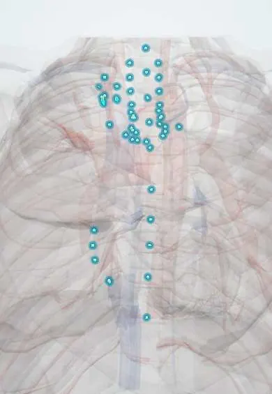
Mediastinal lymph nodes !
The mediastinal lymph nodes sit in the centre of the chest around the airways and heart; they filter lymph from the lungs and chest organs.
Opens in the Durgical app, no sign-up needed.
What it is
Lymph nodes around the trachea, bronchi, heart and esophagus.
What it does
Filter lymph from the lungs and chest organs.
Key facts
- Where
- Anterior mediastinal, paratracheal, tracheobronchial (including subcarinal), bronchopulmonary (hilar), posterior mediastinal
- Drains
- Lungs and bronchi (pulmonary to hilar to tracheobronchial to paratracheal), heart, pericardium, esophagus, thymus
- Into
- Bronchomediastinal trunks, then thoracic duct, right lymphatic duct or venous angles
Relations
- In front
- Prevascular nodes in front of the great vessels and brachiocephalic veins, near the thymus
- Lateral
- Paratracheal nodes on each side of the trachea and hilar nodes at each lung root; on the left, subaortic nodes in the aortopulmonary window beside the left recurrent laryngeal nerve
- Below
- Subcarinal nodes below the tracheal bifurcation, in front of the esophagus and above the left atrium
- Behind
- Paraesophageal and para-aortic nodes in the posterior mediastinum, along the esophagus and descending aorta
How to find it
Not palpable. They are named by numbered stations on CT and PET: 1 low cervical and supraclavicular, 2 and 4 paratracheal, 3 prevascular and retrotracheal, 5 subaortic, 6 para-aortic, 7 subcarinal, 8 paraesophageal, 9 pulmonary ligament and 10 hilar. Stations 2, 4 and 7 are reached by EBUS or mediastinoscopy.
On imaging
On CT a node over about 1 cm in short axis is usually called enlarged; PET-CT adds metabolic activity. On a frontal X-ray, enlargement widens the right paratracheal stripe or fills the aortopulmonary window. Calcified nodes suggest old tuberculosis or fungal infection; eggshell calcification suggests silicosis or sarcoidosis.
Common problems
- Enlarge with infections, sarcoidosis or cancer; checking them is key to staging lung cancer.
How it is diagnosed
CT and PET scans; biopsy through a bronchoscope (EBUS).
Clinical notes
- Sampled by endobronchial ultrasound-guided needle aspiration (EBUS) or by cervical mediastinoscopy; aortopulmonary window nodes need an anterior mediastinotomy or thoracoscopy
- Enlarged aortopulmonary window nodes can compress the left recurrent laryngeal nerve and cause hoarseness
- Tuberculosis enlarges hilar and paratracheal nodes, especially in children, where they can compress a bronchus and collapse a lobe
- Symmetric hilar and right paratracheal enlargement is the classic pattern of sarcoidosis
- Bulky nodes from lymphoma or lung cancer can obstruct the superior vena cava or compress the airway
Related conditions
- HIV infection and AIDS
- Hodgkin lymphoma
- Superior vena cava syndrome
- Castleman disease
- Histoplasmosis
- Anthrax
- Sarcoidosis
- Mediastinal masses (thymoma and anterior mediastinal mass)
- Mediastinitis (acute and fibrosing)
Test yourself
Think of the answer, then tap the question to see it.
In lung cancer staging, what N category is spread to same-side mediastinal or subcarinal nodes, and to opposite-side nodes?
Same-side mediastinal or subcarinal is N2; opposite-side mediastinal or supraclavicular is N3.
Why can a left lower lobe tumour spread to the right mediastinum?
Lymph from the left lower lobe often crosses to the right paratracheal nodes.
What does enlargement of the subcarinal lymph nodes do to the carina?
It widens the carinal angle and can indent the esophagus and the left atrium.
Which mediastinal lymph node pattern is classic for sarcoidosis?
Symmetric hilar and right paratracheal enlargement.
Why can enlarged aortopulmonary window lymph nodes cause hoarseness?
They lie beside the left recurrent laryngeal nerve and can compress it.
Educational information, not medical advice. For any symptom, see a health professional.
Its names
- Official Latin (TA2)
- nodi lymphoidei mediastinales; nodi tracheobronchiales TA2 5254
- Official Spanish (DeCS)
- Nódulos linfáticos mediastínicos DeCS D008198
- English (TA2)
- Mediastinal lymph nodes
Sources: Terminologia Anatomica (TA2); DeCS; Anatomy, Thorax, Mediastinal Lymph Nodes; Tratado de anatomía humana (9.ª ed.), tomo II, Libro IV Angiología, sección 1, cap. I Corazón (arts. VI–IX) y cap. II Pericardio.
Spotted a mistake?We are always improving for the community. Tell us what is wrong and our team will review it.Report a mistake
Representative model, for educational purposes only.