Conditions · Pediatrics and neonatology
Bronchiolitis (RSV) !
A viral chest infection of babies under 2 that inflames the smallest airways, causing cough, wheeze and trouble breathing. RSV is the main cause.
Updated 2026-11-01
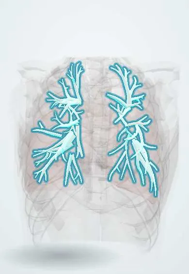
Structures involved
Tap one to see what it is and open it in 3D.
Microorganisms that cause it
In the clinical exam
Why it happens
What it is
Acute viral infection of the lower respiratory tract in children under 2 years, with inflammation and mucus plugging of the bronchioles. It is the first episode of wheezing with respiratory distress after a cold-like start, and respiratory syncytial virus (RSV) causes most cases.
Who gets it
The leading cause of hospital admission in infants worldwide. In Colombia and other tropical countries RSV circulates with peaks during the rainy seasons rather than a single winter. Most severe cases occur in infants under 6 months, including previously healthy babies.
Causes
- Respiratory syncytial virus (RSV), the main cause
- Rhinovirus, human metapneumovirus, parainfluenza, adenovirus, influenza and SARS-CoV-2
- Spread by respiratory droplets and contaminated hands and surfaces
Risk factors
- Age under 3 months
- Prematurity and bronchopulmonary dysplasia
- Haemodynamically significant congenital heart disease
- Immunodeficiency, neuromuscular disease or Down syndrome
- Not being breastfed, exposure to tobacco smoke, crowding and day care attendance
Pathophysiology
The virus infects the bronchiolar epithelium, causing cell necrosis, oedema, mucus production and sloughed cells that plug the very narrow airways of infants. Partial obstruction traps air (hyperinflation) and complete obstruction collapses alveoli (atelectasis), producing ventilation–perfusion mismatch and hypoxaemia. Young infants may also have central apnoea.
Classification
- Mild: feeding well, mild work of breathing, normal oxygen saturation
- Moderate: retractions, reduced feeding, intermittent low saturation
- Severe: marked distress, grunting, apnoea, persistent hypoxaemia or exhaustion
Symptoms and signs
- Runny nose, mild fever and cough for 1–3 days
- Then fast breathing, wheeze and widespread fine crackles
- Chest retractions and nasal flaring
- Poor feeding and fewer wet nappies
- Apnoea, sometimes the first sign in young infants
Diagnosis
- Clinical
- A clinical diagnosis: age under 2, cold-like prodrome, then first episode of wheeze and/or crackles with increased work of breathing. Assessment of respiratory rate, retractions, hydration and pulse oximetry.
- Tests
- Not needed routinely. Viral antigen or PCR testing may be used for isolation and surveillance; blood gas in severe cases.
- Imaging
- Chest X-ray only if severe or atypical; it may show hyperinflation, peribronchial thickening and patchy atelectasis that can be mistaken for pneumonia.
Differential diagnosis
- Viral-induced wheeze or early asthma (recurrent episodes)
- Pneumonia
- Pertussis
- Foreign body aspiration
- Heart failure from congenital heart disease
Goal of treatment
Supportive care: keep oxygenation and hydration adequate until the infection resolves, and identify infants who need hospital or intensive care.
Treatment
- Conservative
- Nasal suctioning with saline, positioning and small frequent feeds; most infants are managed at home.
- Acute
- Supplemental oxygen when saturation is persistently low, nasogastric or IV fluids if unable to feed, and high-flow nasal cannula or CPAP for respiratory failure.
- Follow-up
- Continuous pulse oximetry and respiratory monitoring in admitted infants, especially for apnoea; contact and droplet isolation.
- Medical
- Bronchodilators, corticosteroids and antibiotics are not routinely recommended; hypertonic saline is used in some hospitalised infants.
Complications
- Apnoea, especially in premature or very young infants
- Respiratory failure needing ventilatory support
- Dehydration
- Secondary bacterial infection, such as otitis media
- Recurrent wheezing in later childhood
Prognosis
Most children recover in 1–2 weeks, although cough can last longer. Mortality is low in previously healthy infants but higher in premature babies and those with heart or lung disease.
Prevention
- Handwashing and avoiding contact with people with colds
- Breastfeeding and a smoke-free home
- Long-acting RSV monoclonal antibodies (nirsevimab; palivizumab in selected high-risk infants)
- Maternal RSV vaccination late in pregnancy
Test yourself
Think of the answer, then tap the question to see it.
Which virus causes most cases of bronchiolitis?
Respiratory syncytial virus (RSV).
How is bronchiolitis diagnosed?
Clinically: a child under 2 with a cold-like prodrome followed by a first episode of wheeze or crackles and increased work of breathing.
What is the mainstay of bronchiolitis treatment?
Supportive care: nasal suctioning, hydration and oxygen when saturation is low, with high-flow or CPAP if needed.
Which life-threatening sign can be the first presentation of bronchiolitis in young infants?
Apnoea.
Related conditions
Educational information, not medical advice. For any symptom, see a health professional.
Sources: Bronchiolitis: MedlinePlus Medical Encyclopedia (MedlinePlus, US National Library of Medicine, 2026); Clinical Overview of RSV (Centers for Disease Control and Prevention (CDC), 2025).
Spotted a mistake?We are always improving for the community. Tell us what is wrong and our team will review it.Report a mistake
Representative model, for educational purposes only.